Hysteroscopy: what to expect before, during and after the procedure

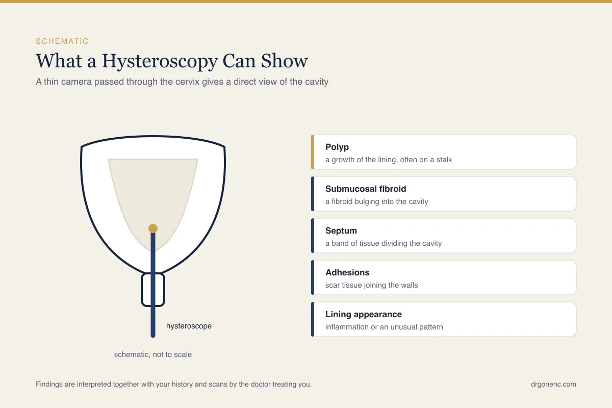
A hysteroscopy is a procedure that lets a doctor look directly inside the uterine cavity, using a thin telescope passed through the cervix. It is one of the few examinations in gynaecology that does not work out what the cavity looks like from a shadow or an outline, but shows the lining itself, the openings of both fallopian tubes, and anything growing or scarring inside. Most people meet the word for the first time when it appears on a request form after a scan, after months of heavy or irregular bleeding, or after a treatment cycle that did not work.
This article explains what a hysteroscopy is and what it can show, the difference between a diagnostic and an operative procedure, what an office or outpatient hysteroscopy involves, the reasons it is requested, when in the menstrual cycle it is usually arranged, how to prepare, what happens step by step, the anaesthesia options, and what recovery normally looks like. It also covers the warning signs that should prompt a call, and the separate question of hysteroscopy before IVF, where the guidelines are reasonably clear but everyday practice between clinics is not. Whether the procedure is right for you is a decision for the doctor examining you.
What is a hysteroscopy, and what can it show?
A hysteroscope is a narrow telescope, rigid or flexible, carrying a light source and a camera that sends a magnified image to a monitor. Diagnostic instruments are slim, commonly around 3 mm to 5 mm in outer diameter. The scope is passed through the vagina and along the cervical canal into the uterine cavity, so there is no incision anywhere. A small amount of sterile saline, often warmed, runs through the scope at the same time, gently separating the front and back walls of the cavity, which otherwise lie flat against each other and cannot be examined. Some units use carbon dioxide gas rather than fluid to open the cavity, although saline is preferred for outpatient work.
Under direct vision the surface of the endometrium can be assessed along with the shape of the cavity. This is how polyps, fibroids bulging inwards, bands of scar tissue, a dividing wall of tissue, retained pregnancy tissue and a displaced contraceptive coil are identified. Both tubal openings can be inspected, and a sample of lining can be taken from a specific area that looks abnormal, rather than blindly from the cavity as a whole. Where treatment is planned, the same access route is used to carry it out.
It is just as important to know what a hysteroscopy does not show. It examines the inside of the cavity only, so it says nothing about the ovaries, nothing about the outer surface of the uterus, and nothing about endometriosis or adhesions elsewhere in the pelvis. It does not measure the thickness of the muscle wall, and on its own it does not establish whether the tubes are open. For those questions, ultrasound, contrast imaging or laparoscopy answer what hysteroscopy cannot, which is why the tests are used alongside one another.
Diagnostic hysteroscopy and operative hysteroscopy
A diagnostic hysteroscopy is an inspection. The aim is to see the cavity, describe what is there, and if necessary take a biopsy. An operative hysteroscopy treats what is found. The instruments differ: an operating hysteroscope has a working channel through which fine scissors, graspers or a bipolar electrode pass, while larger work uses a resectoscope or a mechanical tissue removal device that cuts and suctions tissue at the same time. The optical part of the examination is the same in both cases.
Many units now work on a see and treat basis. With slim instruments, a small polyp can often be removed at the same outpatient visit in which it is found, which spares a second appointment and a second anaesthetic. Larger polyps, fibroids projecting into the cavity, a dividing septum, dense adhesions and retained tissue generally need more operating time, a wider instrument and better pain control, so they are planned as a separate day case procedure rather than attempted in a clinic room.
This distinction matters when you are consented. Before the day you should know which of the two is intended, and what the plan is if something unexpected is found: whether the team will treat it there and then, or stop, explain and rebook. Any tissue removed is sent for pathological examination, and that report usually takes days to a couple of weeks. Which approach suits your situation is a judgement for the doctor who has seen your scans and your history.
Office hysteroscopy and hysteroscopy in an operating theatre
An office hysteroscopy, also called an outpatient hysteroscopy, takes place in a clinic examination room without general anaesthesia. Many units use a vaginoscopic or no touch technique, in which the scope is guided along the vagina and into the cervical canal under vision, with no speculum and no instrument gripping the cervix. This approach tends to be better tolerated, and the RCOG guideline on outpatient hysteroscopy (Green-top Guideline No. 59) recommends it as the standard technique for a diagnostic outpatient procedure. Practice elsewhere varies: ACOG describes a speculum being placed first, which is still routine in some units. You stay awake, can watch the screen, and in most cases walk out shortly afterwards.
A theatre procedure is arranged when more is needed: a longer operating time, wider instruments, a cervical canal that is likely to be difficult, or a previous attempt that was too uncomfortable to complete. It is usually a day case, so you come in and go home the same day, although an overnight stay is occasionally needed after a general anaesthetic or a longer operative procedure. It involves sedation, regional or general anaesthesia and the fasting and escort arrangements that go with them. Neither setting is a lesser version of the other; they answer different questions, and the choice follows what has to be done.
| Feature | Office (outpatient) hysteroscopy | Hysteroscopy in an operating theatre |
|---|---|---|
| Where it is done | A clinic examination room | A day case operating theatre |
| Anaesthesia | None, or a local anaesthetic block | Sedation, regional or general anaesthesia |
| Instrument diameter | Slim, commonly around 3 mm to 5 mm | Often wider, to carry larger working instruments |
| What is usually possible | Inspection, targeted biopsy, removal of a small polyp, retrieval of a coil | Larger polyps, fibroids inside the cavity, division of a septum, release of adhesions |
| Length of the procedure | Usually about 5 to 15 minutes; longer if a biopsy or polyp removal is added | Longer, planned around what is being treated |
| Being awake | Awake; findings can be explained as they appear | Asleep or sedated; findings are explained afterwards |
| Going home | Usually soon afterwards, often able to drive unless inhaled pain relief was used | Usually home the same day, occasionally overnight; an escort is needed and driving is not allowed that day |
Why a hysteroscopy is requested
A hysteroscopy is not a general screening test. It is requested to answer a specific question that imaging has either raised or cannot settle, and the question should be clear before the appointment is made. The most common reasons fall into three groups: an abnormality seen on a scan, a bleeding pattern that needs explaining, and a fertility history that points towards the cavity. The list below covers the situations in which it most often comes up.
- An abnormality on ultrasound, such as a suspected endometrial polyp or a fibroid pressing into the cavity
- Heavy, prolonged or irregular menstrual bleeding, or bleeding between periods
- Bleeding after the menopause, where the lining needs to be seen and sampled
- Suspected scar tissue inside the cavity, often after a previous uterine procedure or infection
- A suspected uterine septum or another congenital variation in the shape of the cavity
- Repeated implantation failure or recurrent pregnancy loss, as one part of a wider assessment
- A contraceptive coil that cannot be located or removed in the usual way
- Pregnancy tissue thought to be retained after a miscarriage or a delivery
- Difficulty passing the catheter at a previous embryo transfer, or a cervical canal that is hard to negotiate
Where the reason is bleeding after the menopause, or a thickened lining on a scan, the purpose of seeing and sampling the endometrium is to rule out hyperplasia or endometrial cancer as well as to find a polyp or a fibroid. Most such samples come back benign. The answer comes from the pathology report rather than from the appearance on the screen, which is why the report matters even when the cavity looked normal, and your own doctor goes through what it means and whether anything further is needed.
Being offered the procedure does not imply that something serious has been found. In many cases the cavity turns out to be normal, and how often that happens depends heavily on why the test was arranged. A normal result is a useful one rather than a wasted one, because it directs attention elsewhere. Equally, a finding on a scan is not by itself a reason to operate. Whether a particular finding needs treating, observing or simply recording is a judgement your own doctor makes with the whole picture in front of them.
What a hysteroscopy adds to an ultrasound scan or an HSG
Transvaginal ultrasound is almost always the first test. It measures the thickness of the lining in millimetres, maps fibroids and their relation to the cavity, and often raises the suspicion of a polyp. Adding fluid to the cavity during the scan, known as saline infusion sonography, outlines the inner surface far better than a plain scan and can distinguish a polyp from a thickened fold. For many people these studies answer the question completely, and no hysteroscopy follows.
A hysterosalpingogram, or HSG, uses contrast and X ray images, and an ultrasound based version uses a foam or bubble contrast instead. These tests show the silhouette of the cavity and whether contrast passes along the tubes, which is their real value. Because they work from an outline, however, the shape they display can be read in more than one way: a septum and a partly divided uterus can produce a similar picture, and a small lesion can be either missed or over called.
Hysteroscopy assesses the inside of the cavity directly, because the surface is seen rather than inferred, and because a biopsy or treatment can follow in the same sitting. It does not replace imaging. The scans describe the muscle wall, the ovaries and the position of fibroids within the wall, none of which the camera can see from inside. Where the outside of the uterus and the pelvis also need assessment, laparoscopy may be arranged at the same time.
When in the menstrual cycle is a hysteroscopy done?
For someone who still has periods, the usual window is the first half of the cycle, after bleeding has finished and before ovulation. In a 28 day cycle that is roughly day 6 to day 12, counting the first day of bleeding as day 1. The reason is practical: at that point the lining is at its thinnest and flattest, so the view is clear, polyps and adhesions stand out, and there is little chance of an early pregnancy being present.
The second half of the cycle is less suitable. After ovulation the lining becomes thick and folded, and those folds can look very like small polyps, which makes the examination harder to interpret. Active bleeding is also unhelpful, because blood and clot obscure the surface and heavy bleeding can defeat the fluid running through the scope. Pregnancy has to be excluded before the procedure goes ahead, which is another reason the early part of the cycle is preferred.
Not everyone has a predictable cycle to plan around. Where bleeding is irregular, where a continuous progestogen or combined hormonal contraception is being taken, or where periods have stopped after the menopause, timing is more flexible and the appointment is set for convenience instead. Urgent indications, such as bleeding after the menopause, are not delayed to chase an ideal day. Your doctor sets the date according to your own pattern and the reason for the examination.
Preparing for a hysteroscopy
Preparation for an outpatient procedure is usually straightforward, and most of it is information rather than restriction. What applies to you depends on the setting and on whether any anaesthesia is planned, so the instructions from your own unit take precedence over any general list. The points below are the ones that come up most often, and each of them is worth confirming when the appointment is booked rather than on the day.
- Tell the team every medicine you take, including blood thinning medication, and mention any allergies
- Say in advance if you have painful periods that make you faint, if previous vaginal examinations or smear tests were painful, or if you have experienced sexual violence, since any of these can change the setting and the anaesthesia that is offered, and it is far easier to arrange before the day than on it
- Confirm whether to eat and drink beforehand; fasting applies only when sedation or general anaesthesia is arranged
- Use reliable contraception, or avoid intercourse between your last period and the appointment, since you may be asked to do a urine pregnancy test when you arrive
- Ask whether to take a simple painkiller such as paracetamol or a non-steroidal anti-inflammatory about an hour before an outpatient procedure
- Arrange someone to take you home if sedation or general anaesthesia is planned
- Bring a sanitary pad, since light bleeding afterwards is usual
- Ask whether treatment may be carried out at the same visit, so that you are consented for it in advance
Two forms of preparation are sometimes used when the cervical canal is expected to be tight, for example after the menopause, after cervical surgery or where a previous attempt failed: a short course of vaginal oestrogen, or a prostaglandin such as misoprostol given before the procedure. Neither is routine, and units differ in whether they use them at all. Antibiotics are not given as a matter of course for a straightforward examination. Whether any of this applies to you is for your doctor to decide.
What happens during a hysteroscopy
You will be asked to empty your bladder, change, and lie on a couch with your legs supported, in the position used for a cervical screening test. Consent is confirmed and the steps are explained again. If a vaginoscopic technique is used, no speculum is placed; otherwise a speculum is inserted first and the cervix is cleaned. Where a local anaesthetic has been agreed, it is applied to the cervix or injected around it at this point and given a few minutes to work.
The scope is then guided along the cervical canal under direct vision while saline flows ahead of it to open the way. Most people feel pressure and cramping that build as the cavity fills, strongest while the scope passes through the canal. Fluid running back out is expected, and the sensation of needing to pass water is common. Less often there is a wave of nausea, sweating and light headedness. Where carbon dioxide rather than saline is used to open the cavity, discomfort at the tip of the shoulder can also occur. Any of these should be reported straight away rather than endured.
Once inside, the examination is systematic: the whole cavity, the front and back walls, the fundus, and both tubal openings. If you are awake, the findings can be described to you as they appear on the screen, and most people find it easier to follow the image than to look away. A targeted biopsy is taken if the lining needs sampling. Where treatment was planned and is feasible, it is carried out now, with the instruments passed down the working channel.
At the end the scope is withdrawn, the fluid drains, and the cramping usually eases within minutes. A purely diagnostic inspection usually takes about 5 to 15 minutes, and longer if a biopsy is taken or a polyp removed, although the appointment as a whole is considerably longer because of consent, positioning and a short period of observation afterwards. You will be told what was seen before you leave, and if tissue was removed you will be given an idea of when the pathology report is expected and how the result will reach you.
Anaesthesia, discomfort and pain relief
There are several options, and they are chosen rather than assumed. A diagnostic outpatient procedure may be done with no anaesthesia at all, or with a local anaesthetic applied to the cervix as a gel or spray, or injected around it as a paracervical or intracervical block using an agent such as lidocaine. Some units also offer inhaled pain relief during the procedure: gas and air, which is a mixture of nitrous oxide and oxygen breathed through a mouthpiece and taken as needed, or in a few hospitals methoxyflurane. Both act within a minute or two and wear off quickly, although you may be asked to wait longer in the unit before driving if you use them. Conscious sedation is a further intermediate step. Regional or general anaesthesia is used for operative work, for a difficult cervix, or where the alternative would not be tolerated.
It is worth being plain about sensation, because vague reassurance helps nobody. Most people describe cramping like a strong period pain, building while the cavity is distended and settling quickly once the scope is removed. Some feel very little and are surprised how quick it was. Others find it genuinely painful, and that is not a failure of preparation or of nerve. Discomfort tends to be greater where the cervix is narrow or scarred, where no vaginal birth has taken place, where the procedure runs long, or where a previous examination was painful.
Several things appear to reduce it. A no touch vaginoscopic approach and a smaller diameter scope are the measures supported by randomised trials. A steady unhurried technique, a running commentary and warming the fluid are widely used and many patients prefer them, although the trial evidence for them is less consistent. A simple painkiller taken beforehand, if your doctor agrees it suits you, is reasonable. Most importantly, you can ask for the procedure to stop at any moment, and asking is not a problem for the team: an examination that is too uncomfortable can be abandoned and rearranged under anaesthesia. Agree that plan in advance, so it does not have to be negotiated during the procedure.
Recovery, what is normal, and the warning signs
After an outpatient procedure most people rest for a few minutes and then go home, often driving themselves. Cramping similar to period pain is usual for a few hours and sometimes for a day or two, and simple painkillers are normally enough. Light bleeding or spotting is expected for several days and occasionally for about a week, and a watery pink discharge from the saline is common in the first day. Normal activity, including work, can usually be resumed the same day or the next, but hold off on exercise, tampons and intercourse until the pain and bleeding have stopped, commonly about a week.
Recovery after sedation or general anaesthesia takes longer. Drowsiness lasts for the rest of that day, so driving, alcohol and important decisions are ruled out, and someone should be with you. After a straightforward day case procedure many people return to their usual routine the following day, while recovery after extensive operative work inside the cavity takes longer. Follow the instructions your own team gives you.
- Heavy bleeding, such as soaking through a pad within an hour, or passing large clots
- A temperature, shivering, or feeling generally unwell in the days afterwards
- Vaginal discharge with an unpleasant smell
- Pain that is severe, getting worse, or not relieved by the painkillers you were advised to take
- Fainting, breathlessness or chest pain
- Pain together with vomiting, or an abdomen that becomes swollen and tender
Complications are uncommon, and most hysteroscopies pass without any. The recognised ones are failure to enter the cavity through a tight cervix, a vasovagal reaction with faintness during the procedure, infection, a small perforation of the uterine wall, bleeding, absorption of too much distension fluid during longer operative procedures, and new intrauterine adhesions after extensive surgery inside the cavity. Perforation of the uterine wall is rare in a diagnostic outpatient procedure and becomes a little more likely when a polyp or fibroid is removed at the same visit, and infection is uncommon. Perforation usually needs nothing more than observation, occasionally an overnight stay. Units take specific steps to limit each of these. Any of the symptoms listed above should be discussed with the team who carried out the procedure rather than left to settle.
Hysteroscopy before IVF: when it is suggested and when it is not
The cavity is where an embryo has to implant, so its condition is reasonably part of the preparation for treatment. A polyp, a fibroid pressing inwards, a septum, retained tissue or a band of scar tissue can all change what is planned, and are among the factors considered when a cycle has not worked. That is the thinking behind discussing why an IVF cycle fails in terms of the uterus as well as the embryo.
Where practice genuinely differs is over routine use. For someone with no symptoms and a normal transvaginal ultrasound, NICE is explicit on this point: its 2026 fertility guideline (NG257) recommends not offering hysteroscopy unless a uterine or endometrial abnormality is clinically suspected (recommendation 1.18.16), and separately recommends not offering hysteroscopy as a pre-treatment to improve the outcome of IVF (recommendation 1.40.2). That applies to repeat cycles as well as a first one. Two randomised trials published in 2016, one in women starting a first cycle and one in women with two or more previous unsuccessful cycles, found no improvement in live birth from inspecting a cavity that looked normal on ultrasound. Some centres nonetheless offer it routinely, and patients are often told conflicting things as a result.
Agreement is much wider in other situations. Hysteroscopy is generally accepted where a scan or saline sonography suggests a lesion, where previous surgery or infection makes adhesions likely, where a septum is suspected, where tissue may have been retained, and where a previous transfer was technically difficult, since the cervical canal can be assessed at the same visit. After repeated implantation failure some units also take an endometrial biopsy to look for chronic endometritis. This one is contested rather than agreed: NICE recommends against testing the endometrium for infectious chronic endometritis as an add-on before embryo transfer (recommendation 1.41.1), and whether it is worth doing in your case is a decision for your own clinic.
Timing is the last question. A diagnostic procedure is usually arranged before stimulation begins, or in a cycle preceding a frozen transfer, so that the result is available while the plan is still open. Where something has been treated inside the cavity, an interval is normally allowed for the lining to heal before transfer, and its length depends on how much was done. Some units also check the cavity again after extensive surgery before proceeding.
None of this translates directly from one person to another. Whether a hysteroscopy adds anything in your case depends on your scans, your bleeding pattern, any previous uterine surgery and what happened in earlier treatment cycles, and the balance of those factors is not something a general article can settle. The assessment is individual, and the decision to recommend or to leave out the procedure belongs to the physician treating you.
Related Reading
- Uterine Septum and Fertility: What to Know
- Asherman Syndrome (Intrauterine Adhesions) and Fertility
- Chronic Endometritis and How It Affects Implantation
- Endometrial Lining and Implantation: What Matters
- Why Does IVF Fail? Common Reasons and What to Do Next
- Uterine polyps and fertility: endometrial polyps, symptoms and removal
- Uterine fibroids and fertility: why the position of a fibroid in the uterus matters more than its size
Sources and references
This article is supported by the following independent, authoritative sources:
- NHS (UK), Hysteroscopy
- ACOG (American College of Obstetricians and Gynecologists), Hysteroscopy
- RCOG (Royal College of Obstetricians and Gynaecologists), Outpatient hysteroscopy
- RCOG, Outpatient Hysteroscopy (Green-top Guideline No. 59)
- NICE (UK), Fertility problems: assessment and treatment (NG257)
- NICE (UK), NG257, Procedures used during in vitro fertilisation (IVF)