Uterine polyps and fertility: endometrial polyps, symptoms and removal

An endometrial polyp is a small, localised overgrowth of the lining of the womb that grows into the uterine cavity. Polyps are among the more frequent findings on a pelvic scan, and seeing the word on a report can be unsettling, particularly for someone who is trying to conceive. In most cases a polyp is a benign, slow growing piece of tissue rather than a sign of anything sinister, and in a great many women it causes no symptoms whatsoever.
This article explains what a polyp is made of, how it differs from a fibroid, the bleeding patterns it can produce, how it is identified on ultrasound and at hysteroscopy, and why its effect on implantation is still discussed rather than settled. It also covers when removal is advised, what a hysteroscopic polypectomy involves, how recovery usually goes, whether polyps return, and what happens when one is found before or during an IVF programme. Everything here is general information. Your own scan, your own bleeding history and any decision about surgery belong to the doctor who has examined you.
What an endometrial polyp is
A polyp is a focal outgrowth of the endometrium, the lining that is built up and shed with each menstrual cycle. Under the microscope it contains endometrial glands, supporting stromal tissue and a central core of blood vessels that feeds it. Some polyps sit on a narrow stalk, described as pedunculated, and can move within the cavity or even reach down through the cervix. Others have a broad base, sit flat against the wall and are described as sessile.
Sizes vary widely. Many are only a few millimetres across and are noticed only because a scan was being done for another reason. Others reach three or four centimetres and fill much of the cavity. A woman may have a single polyp or several, and multiple small polyps behave differently from one large one when it comes to both bleeding and treatment decisions. Position matters too, since a polyp near a tubal opening raises different questions from one on the front wall.
It is worth separating a few terms that often appear on the same report. An endometrial polyp sits inside the uterine cavity. A cervical polyp grows from the canal of the cervix and is frequently visible at a routine examination. Endometrial hyperplasia is a generalised thickening of the whole lining rather than a discrete lump. And a focal area of thickening seen on a single scan is not automatically a polyp, which is one reason the scan is often repeated at a different point in the cycle.
Why polyps develop, and who is more often affected
Polyp tissue is sensitive to oestrogen, and the usual explanation is that a small area of lining responds more strongly to hormonal signals than the tissue around it and keeps growing while the rest of the lining is shed. That is why polyps are found more often in situations where oestrogen exposure is relatively unopposed by progesterone, and why they are seen more frequently as women move through their forties and towards the menopause.
- Increasing age, with polyps found more often in the years around and after the menopause.
- Carrying significant excess weight, since fat tissue converts other hormones into oestrogen.
- Cycles in which ovulation is infrequent, so the lining meets oestrogen without a regular progesterone phase.
- Treatment with tamoxifen for breast cancer, which acts on the endometrium in an oestrogen like way.
- Oestrogen therapy after the menopause taken without adequate progestogen cover.
- Raised blood pressure, which appears in several published series although the link is less firmly established.
None of this means a polyp is anyone's fault, and most women who develop one have no identifiable risk factor at all. Equally, having several of these features does not mean a polyp will form. The practical value of the list lies elsewhere: it helps your doctor judge how carefully a particular polyp needs to be investigated, because the same features that favour polyp growth also influence how likely the tissue is to show any abnormal change.
Endometrial polyp or fibroid?
Polyps and fibroids are frequently confused, partly because both can bulge into the cavity and cause heavy or irregular bleeding, and partly because reports sometimes use the vague word growth for either. They arise from different tissues, behave differently and are treated differently. A fibroid, properly called a leiomyoma, is a firm ball of smooth muscle arising in the muscular wall of the uterus. A polyp is soft tissue arising from the lining that covers that wall.
| Feature | Endometrial polyp | Uterine fibroid |
|---|---|---|
| Tissue of origin | Glands and supporting tissue of the womb lining (endometrium) | Smooth muscle of the uterine wall (myometrium) |
| Consistency | Soft and fleshy | Firm and rubbery |
| Usual size range | Most often a few millimetres to about two centimetres, occasionally three or four | A few millimetres to many centimetres |
| Where it sits | Projects into the cavity, often on a narrow stalk | Within the muscle wall, bulging into the cavity or outwards |
| Bleeding pattern most often reported | Spotting between periods, prolonged periods, unpredictable loss | Heavy menstrual bleeding, with pressure and urinary symptoms when large |
| Imaging used | Transvaginal ultrasound, saline infusion sonography, hysteroscopy | Transvaginal ultrasound, magnetic resonance imaging when mapping is needed |
| How the diagnosis is confirmed | Histology of the polyp once it has been removed | Imaging is usually sufficient, histology if it is removed |
| Usual procedure | Hysteroscopic polypectomy | Myomectomy: hysteroscopic, laparoscopic or open according to position |
| Can new ones form later | Yes, further polyps can develop | Yes, further fibroids can develop |
The distinction matters for the plan rather than for the label alone. A soft polyp within the cavity can usually be dealt with in a single short hysteroscopic procedure. A fibroid may be managed by observation, by medication, or by surgery through the cavity, through the abdomen with keyhole instruments or through an open incision, depending on how deeply it sits in the muscle. Where imaging cannot separate the two with confidence, hysteroscopy or magnetic resonance imaging is sometimes added before anything is decided.
Symptoms, and why many polyps cause none
The commonest reason a polyp is found is that nothing at all pointed to it. A scan performed for pelvic pain, for a fertility assessment or as part of routine gynaecological care shows a small mass within the cavity, and the woman has noticed no change in her periods. Silent polyps are ordinary rather than unusual, especially when they are small. When symptoms do appear, they are almost always about bleeding rather than pain.
- Spotting or light bleeding between periods.
- Periods that last longer than they used to, or that trail off with several days of brown loss.
- Heavier menstrual bleeding, sometimes with clots.
- Bleeding or spotting after intercourse.
- A watery or blood stained discharge outside the time of a period.
- Any bleeding at all after the menopause, which always needs assessment rather than watching.
- Difficulty conceiving, or repeated early pregnancy loss, with no other explanation found, although the strength of this link is still debated and is discussed further below.
The bleeding comes from the polyp's own fragile surface vessels, which are not under the same hormonal control as the rest of the lining and can bleed at any point in the cycle. That is why the pattern is often described as unpredictable rather than simply heavy. A polyp sitting on a long stalk that reaches the cervix can also bleed after intercourse, for the straightforward reason that it is exposed to contact.
Pain is not a typical feature. Some women describe cramping as a stalked polyp is pushed towards the cervix, which can feel like a period pain outside the expected days, but a polyp does not usually cause the pressure sensation, bloating or urinary frequency that a large fibroid produces. If you have significant pain, it is worth asking whether something else is also present rather than assuming the polyp explains everything.
How a polyp is found: ultrasound, saline infusion sonography and hysteroscopy
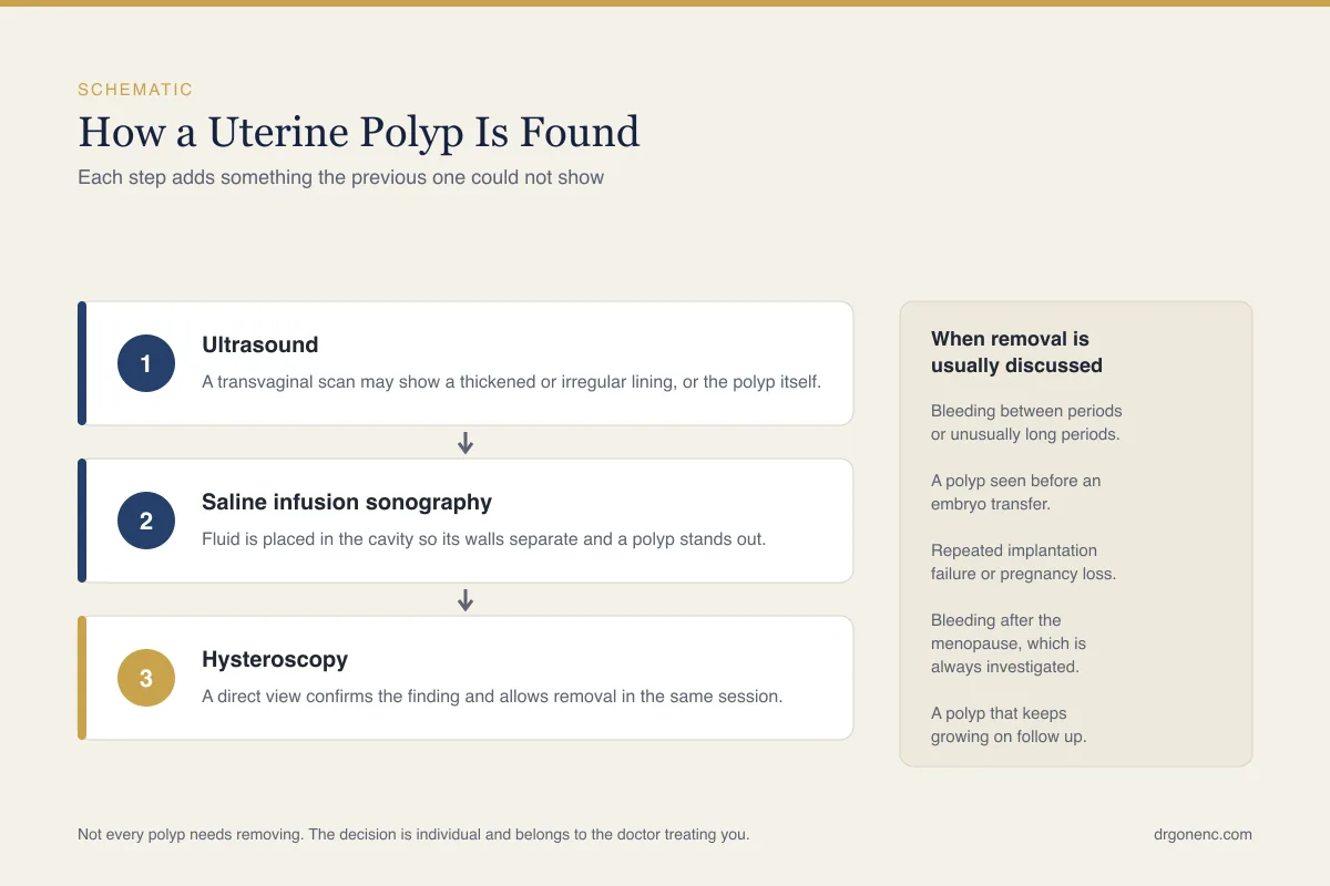
Transvaginal ultrasound is the first test. A polyp typically appears as a bright, rounded area within the cavity, and colour Doppler often shows a single feeding vessel running into it, which is a helpful sign. Timing makes a real difference: scanning in the first half of the cycle, soon after a period when the lining is thin, makes a discrete polyp far easier to see. Scanning in the second half, when the lining is thick, can both hide a polyp and create a false impression of one.
Saline infusion sonography, also written as saline infusion sonohysterography or SIS, refines the picture. A fine catheter is passed through the cervix and a small volume of sterile saline is instilled to separate the front and back walls of the cavity while the scan continues. Against the dark fluid a polyp stands out as a projection with a stalk, and the examination also shows how much of the cavity it occupies. It is done in the clinic and takes only a few minutes.
Hysteroscopy, in which a thin telescope is passed through the cervix so that the cavity is viewed directly, is the reference standard. It shows the number, size, position and surface appearance of any polyps, and it allows removal at the same sitting. Many units now work this way, assessing and treating in one visit where that is appropriate and has been agreed in advance. A hysteroscopy appointment itself, including how it is carried out and how it feels, is described separately.
Two points cause recurring confusion. A blind dilatation and curettage, in which the lining is scraped without a camera, misses polyps regularly and is no longer regarded as an adequate way to remove one. And an endometrial biopsy taken with a fine sampling device reports on the lining in general rather than on a particular polyp, so a normal biopsy neither excludes a polyp nor confirms that a polyp seen on a scan is harmless.
Polyps and implantation: why the effect is still discussed
There are sound reasons to think a polyp can interfere with implantation. It occupies part of the cavity where an embryo needs to settle, it may sit across a tubal opening and hinder the passage of sperm or egg, and the lining over and around it does not behave normally. Studies of endometrial tissue near polyps have reported changes in the molecular signals associated with receptivity, along with a tendency towards local inflammation.
Polyps also keep company with other findings. Inflammation of the lining is identified more often in women who have polyps than in those who do not, which matters because inflammation has its own relationship with implantation; our article on chronic endometritis and implantation covers that ground. The thickness and quality of the surrounding lining is relevant in much the same way, and is discussed in our article on endometrial lining and implantation.
Where the picture becomes less clear is in what happens after removal. Removing a polyp before intrauterine insemination has been examined in a single small randomised trial whose findings favoured removal, and that trial has not since been repeated. For women with otherwise unexplained infertility, and for small polyps found by chance in women with no symptoms, the evidence is largely observational, the polyps studied differ widely in size and number, and professional bodies have been careful not to convert a plausible mechanism into a routine operation for everybody.
A reasonable way to hold all this is that a polyp is a correctable finding with a believable mechanism, not an established cause in any individual case. Where a couple has had repeated unsuccessful transfers or losses, the balance usually tips towards removal; our article on why IVF cycles fail sets it alongside the other factors that are examined. Where a tiny polyp turns up in a woman who has conceived easily before, watching is defensible. Your doctor weighs these together.
The cancer question, answered plainly
Almost everyone who is told they have a polyp wonders about cancer, and the honest answer is reassuring without being absolute. The large majority of endometrial polyps are entirely benign. A minority contain areas of hyperplasia, meaning overgrown lining, and a smaller minority again contain atypical cells or, rarely, a cancer. The likelihood is low, and lower still in a woman who has not yet reached the menopause and has no abnormal bleeding.
Certain features raise the level of concern and change how promptly a polyp is dealt with: bleeding after the menopause, bleeding at any age that is new or persistent, a larger polyp, being postmenopausal, treatment with tamoxifen, significant excess weight, and a family history suggesting Lynch syndrome. None of these is a diagnosis in itself. They simply mean that the sensible course is to remove the tissue and examine it rather than to watch and wait.
This is why removed polyps are sent for histological examination as a matter of routine, even when everything looked ordinary down the telescope. The microscope settles the question and imaging cannot. If a report describes a benign endometrial polyp, that is a reliable answer about the tissue that was removed. If it describes hyperplasia, with or without atypia, the conversation moves on to how the lining as a whole should be managed, which is a separate discussion with its own plan.
For a woman in her twenties or thirties with regular periods whose polyp was found during a fertility scan, the conversation is usually about implantation rather than about cancer. For a woman who has bled after the menopause it is the other way round, and the assessment should not be delayed while other matters are arranged. Both of those conversations deserve a direct answer from the clinician who knows your history and has read your images.
When removal is advised, and when observation is reasonable
There is no single rule, and the decision comes from the combination of symptoms, age, menopausal status, the polyp itself and what you are trying to achieve. Guidance from different bodies emphasises different parts of that picture. AAGL guidance on the diagnosis and management of endometrial polyps takes the view that removal may reasonably be considered in women with infertility, while ESHRE, reviewing the same question for unexplained infertility and for recurrent implantation failure, found the evidence too limited to recommend removal routinely for a polyp that was found by chance and is causing no symptoms. NICE, for its part, frames polyps around abnormal bleeding rather than around fertility. That is why two careful doctors may advise differently about the same small, silent polyp. What they tend to agree on is the set of situations in which removal is usually advised.
Removal is generally advised when there is abnormal bleeding that the polyp could explain, when there has been any bleeding after the menopause, when the polyp is large or there are several of them, when features that raise concern about the tissue are present, and when a woman is about to begin fertility treatment and the polyp lies within the cavity where an embryo must implant. In those settings the procedure treats the symptom and answers the tissue question at once.
Observation is reasonable in a narrower set of circumstances: a small polyp, in a woman who has not reached the menopause, with normal periods, no bleeding between them, no features of concern and no immediate plan for treatment. Small polyps can disappear by themselves, which is well documented and more likely with those under about 10 mm, so a repeat scan after a few months is often more informative than proceeding straight to surgery.
It is worth saying plainly what does not work. No tablet, herbal preparation or hormonal treatment reliably removes a polyp. Hormonal medication, including a progestogen releasing intrauterine system, can control bleeding and is sometimes used for that purpose, particularly alongside treatment of the lining as a whole, but the polyp itself is a piece of tissue and removing it means removing it. Whether that is needed in your case is a judgement for your own doctor.
Hysteroscopic polypectomy and what recovery involves
A hysteroscopic polypectomy is performed through the cervix, with no incision anywhere on the abdomen. The hysteroscope is passed into the cavity, the cavity is gently distended with fluid, and the polyp is taken out at its base using fine scissors and grasping forceps, a small mechanical tissue removal device, or a thin electrical loop. Removing the stalk flush with the wall matters more than simply detaching the visible lump, because tissue left at the base can regrow.
Where it is done depends on the polyp and on you. A small polyp is often dealt with in an outpatient or office setting with no anaesthetic, or with local anaesthetic placed around the cervix, and you walk out afterwards. Larger or multiple polyps, a narrow cervix, or simply a wish not to be awake, lead instead to a day case procedure under sedation or general anaesthesia. Discomfort during an outpatient procedure is common and usually cramp like, and it should be discussed beforehand rather than endured in silence.
- Cramping similar to period pain for a day or two, usually eased by simple analgesia such as paracetamol or ibuprofen where that is suitable for you.
- Light bleeding or brown spotting for a few days, sometimes continuing on and off for a week or two.
- A return to normal activity the same day after an outpatient procedure, and usually the next day after sedation or a general anaesthetic. After sedation or a general anaesthetic you should not drive for at least 24 hours and will need someone to take you home.
- Advice from your own unit about tampons, swimming and intercourse until the bleeding has settled, since local practice varies.
- A histology result, usually available within a couple of weeks, and a plan made once it is back.
Contact the unit rather than waiting if you develop bleeding heavier than a period, a fever, an offensive smelling discharge, or pain that is worsening instead of easing. These events are uncommon, but infection, rarely a small perforation of the uterine wall, and rarely scarring inside the cavity are recognised complications of any procedure inside the uterus; our article on Asherman syndrome and intrauterine adhesions explains that last one, which is one reason a focused removal at the base is preferred to a wide scrape. Reporting them early is what allows them to be dealt with promptly. Written instructions and a telephone number should form part of your discharge information.
Recurrence and follow up after a polypectomy
Polyps can come back, and it is better to know that in advance than to be taken by surprise. Recurrence is more likely when there were several polyps rather than one, when the hormonal pattern that encouraged the first one has not changed, and when the original removal was incomplete. It is less likely after a careful hysteroscopic resection at the base than after a blind curettage, which is one of the arguments for working under direct vision.
Routine repeat hysteroscopy for everybody is not standard practice. For most women the plan after a polypectomy is simple: note how the bleeding behaves over the next few cycles, and arrange a scan if the old pattern returns. For women in fertility treatment a scan before the transfer cycle is usual in any case, which provides a natural check. For women taking tamoxifen, or whose histology showed hyperplasia, follow up is individualised and arranged by the team looking after that condition.
If a polyp does recur, the approach is the same as the first time, with one addition: the reason behind it receives more attention. That may mean looking again at weight, at whether ovulation is happening regularly, at any hormone therapy being taken, and at whether the lining as a whole rather than one lump within it is what actually needs treating.
Polyps found before or during an IVF programme
In fertility treatment, polyps usually come to light at one of two moments. The first is the baseline assessment, where the cavity is checked before stimulation begins. A polyp found then is normally removed before the programme starts, because the procedure is short, recovery is quick and it is simpler to begin with a clear cavity than to invest a cycle and wonder afterwards. Most units allow stimulation to begin after the next period, once the lining has regenerated.
The second moment is during stimulation itself, when oestrogen levels rise, the lining thickens and something previously invisible becomes apparent. Caution is needed in both directions here. A thick lining in stimulation can produce an appearance that gets reported as a polyp but proves to be a fold of lining or a small collection of blood, so the finding is often rechecked. Equally, a genuine polyp seen at this stage does not have to bring the cycle to an end.
Where a clear polyp is found late in a stimulation cycle, a common solution is to continue to egg collection, fertilise and freeze the embryos, remove the polyp in a short procedure afterwards, and transfer in a later frozen cycle into a cavity that has been seen to be clear. This separates the two problems instead of forcing a choice between them, and frozen transfer is now a routine part of treatment rather than a fallback position.
One question comes up repeatedly: should everyone have a hysteroscopy before IVF, simply in case? Randomised trials in women whose ultrasound was normal did not show improved live birth after routine hysteroscopy, so most guidance reserves it for a reason, such as an abnormal scan, repeated failed transfers or a cavity that looks suspicious. Assessment here is genuinely individual. What is right for your cavity, your history and your treatment plan is a decision for the physician who examines you and reads your own images.
Related Reading
- Uterine Septum and Fertility: What to Know
- Endometrial Lining and Implantation: What Matters
- Chronic Endometritis and How It Affects Implantation
- Recurrent Miscarriage and Implantation Failure: Causes and What Can Help
- Why Does IVF Fail? Common Reasons and What to Do Next
Sources and references
This article is supported by the following independent, authoritative sources: