Uterine fibroids and fertility: why the position of a fibroid in the uterus matters more than its size

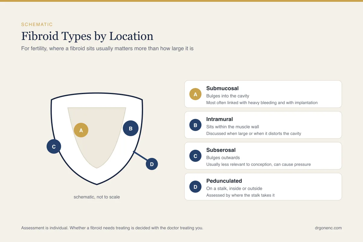
Few findings on a pelvic scan cause as much alarm, and as much unnecessary alarm, as the word fibroid. They are benign growths of the muscle of the uterus, they are among the commonest findings in women of reproductive age, and in the majority of cases they sit quietly in the wall of the uterus without causing symptoms or difficulty conceiving. A smaller group of them genuinely matters, and what separates the two is less about how large the fibroid measures than about where in the uterus it sits and whether it reaches the cavity where an embryo has to implant.
This article sets out what fibroids are and how common they are, the classification that actually guides decisions in fertility care, why a small fibroid bulging into the cavity can count for more than a large one on the outer surface, the symptoms that bring them to attention, how they are assessed, the mechanisms by which they are thought to interfere with conception and implantation, the treatment options including myomectomy, the situations in which watchful waiting is entirely reasonable, how fibroids behave in pregnancy, and what is weighed when a fibroid is found before an IVF cycle. Where bodies genuinely disagree, that is said rather than smoothed over. How any of it applies to a particular uterus is a question for the doctor who has seen the scans.
What a fibroid actually is
A fibroid is a firm, rounded growth made of the same smooth muscle that forms the wall of the uterus, bound together with a dense fibrous matrix. The medical names are leiomyoma and myoma, and the three words describe the same thing. Each fibroid arises from a single muscle cell that has begun to multiply, which is why one uterus can contain several fibroids of quite different sizes, each growing at its own pace and each with its own behaviour over the years.
Fibroids are responsive to oestrogen and progesterone, and that explains most of their natural history. They appear during the reproductive years, rarely before the first period, often enlarge while hormone levels are high, and usually become smaller and less troublesome after the menopause. Growth is not steady or predictable. A fibroid may stay the same size for years, enlarge over a few months, or stop growing entirely, and a single measurement on one scan says little about what it will do next.
It is worth saying plainly what a fibroid is not. It is not a cancer, and the malignant tumour of uterine muscle, leiomyosarcoma, is a separate and uncommon condition rather than the end point of an untreated fibroid. A fibroid is also not an endometrial polyp, which arises from the lining rather than the muscle, and it is not adenomyosis, in which endometrial tissue grows within the muscle wall itself. The three can coexist and can look similar on an ordinary scan, which is one reason imaging is sometimes repeated or extended.
How common fibroids are, and who tends to develop them
Fibroids are the commonest benign growth of the uterus, and far commoner than most people expect: the cumulative incidence across the reproductive years, as the ASRM guideline sets it out, is high enough that most women will have developed at least one by the time they reach the menopause. Most of those women never know it, because the fibroid is found incidentally during a scan arranged for another reason or during routine antenatal imaging. Only a minority ever produce symptoms that need treatment, and only a minority of that minority are in a position where fertility is affected. A diagnosis of fibroids on its own is therefore not a diagnosis of a fertility problem.
The exact cause is not known, and the patient information from both the NHS and ACOG says so plainly. The current understanding is that a fibroid begins when a single muscle cell of the uterus acquires a change that allows it to multiply, and that oestrogen and progesterone then drive how fast it grows rather than start it off, which is why fibroids enlarge in the reproductive years and shrink after the menopause without hormones being the cause. Several factors are associated with developing them. Risk rises with age through the reproductive years, and a family history makes fibroids more likely. Women of Black African ancestry tend to develop them earlier, in greater numbers and at larger sizes, and this difference is consistent enough that it is taken into account when symptoms are assessed; UK guidance also lists Asian background as a factor. An early first period, never having carried a pregnancy to term, a higher body weight, high blood pressure or diabetes, and low vitamin D levels have all been linked with a greater likelihood, while having carried a pregnancy appears to be associated with a lower one.
These associations describe populations rather than people. Many women with every listed risk factor never develop a fibroid, and many with none develop several. In a fertility consultation the figures matter far less than what the scan actually shows: how many fibroids there are, where each one sits in relation to the cavity, what the lining looks like and whether the symptoms described fit the findings. Everything useful follows from those answers rather than from how common fibroids are in general.
The classification that guides fertility decisions
Fibroids are described by their position in the wall of the uterus, and the system in general use is the FIGO numbering, which runs from type 0 to type 8. The numbers sort fibroids into three recognisable families. Submucosal fibroids reach or distort the cavity. Intramural fibroids sit within the muscle wall. Subserosal fibroids project outwards from the outer surface. A ninth category covers unusual positions such as the cervix. The numbering is useful precisely because it records the one thing that drives fertility decisions, which is the relationship of the fibroid to the lining.
A fibroid that spans two compartments is given two numbers, written with a hyphen, the first describing its relationship to the cavity and the second its relationship to the outer surface. A report that says only that there is a fibroid measuring a certain number of centimetres has left out the information a fertility specialist most needs. If the report in hand does not state the type or at least the distance from the cavity, that is a reasonable thing to ask about rather than something to assume.
| Type | Where it sits | FIGO numbering | Relevance to fertility and implantation |
|---|---|---|---|
| Submucosal, pedunculated inside the cavity | Hanging into the cavity on a stalk, covered only by endometrium | Type 0 | The group most consistently linked with heavy bleeding, with difficulty conceiving and with early pregnancy loss. Usually removable through the cervix at hysteroscopy, and removal before fertility treatment is widely advised |
| Submucosal, mostly within the cavity | Bulging into the cavity, with the smaller part of its bulk inside the muscle wall | Type 1 | Treated in practice much like type 0. The cavity is distorted and the endometrium over the fibroid is thinned, so hysteroscopic removal is commonly recommended when pregnancy is the aim |
| Submucosal, mostly within the wall | Touching and indenting the cavity, with the greater part of its bulk inside the muscle wall | Type 2 | Still counted as submucosal and still thought to matter. Removal is technically harder, sometimes needs two sittings, and the risk of thinning the wall is weighed before surgery is offered |
| Intramural, abutting the endometrium | Entirely within the muscle wall but in contact with the lining, without indenting it | Type 3 | The genuinely debated group. Some bodies group it with submucosal fibroids because of its contact with the lining, others with intramural fibroids. Decisions here are individual |
| Intramural | Entirely within the muscle wall, not reaching either the cavity or the outer surface | Type 4 | Evidence is mixed and guidance differs. Larger ones and those close to the lining attract more concern, but a clear benefit from removing a fibroid that is not distorting the cavity is not established |
| Subserosal, mostly within the wall | Bulging outwards, with the greater part of its bulk still inside the muscle wall | Type 5 | Generally not thought to interfere with implantation. May still cause pressure symptoms, and a fibroid in this position can occasionally sit near a tubal opening |
| Subserosal, mostly outside the wall | Projecting from the outer surface of the uterus | Type 6 | Not usually considered a fertility problem. Treatment is driven by symptoms such as pressure, pain or abdominal distension rather than by conception |
| Subserosal, pedunculated | Attached to the outer surface by a stalk, sitting free in the pelvis | Type 7 | Unlikely to affect the cavity at all. The stalk can twist and cause sudden pain, which is a reason for review rather than a fertility concern |
| Other positions | Cervical fibroids, fibroids within the broad ligament, and fibroids that have taken a blood supply from elsewhere | Type 8 | Judged case by case. A cervical fibroid can obstruct the passage of a catheter at embryo transfer or the cervix in labour, so its position is noted before treatment is planned |
Reading across the table, the pattern is consistent. The nearer a fibroid comes to the cavity, the more likely it is to be thought relevant, and the further out it sits, the more the decision shifts from fertility towards symptom relief. The middle of the table is where honest disagreement lives, and where the judgement of the doctor who knows the whole picture, including age, ovarian reserve, previous pregnancies and how long conception has been attempted, carries more weight than any classification on its own.
Why location matters more than size
The question patients most often ask is how big a fibroid has to be before it causes trouble, and the honest answer is that size is the second question rather than the first. A fibroid of a centimetre or two sitting inside the cavity changes the surface on which an embryo has to implant, thins the endometrium that covers it and alters the local blood supply. A fibroid of eight or nine centimetres growing outwards from the back of the uterus may leave the cavity entirely undisturbed. The two findings are not comparable simply because one number is larger.
The mechanism makes this easier to accept. Implantation depends on a receptive endometrium lying against a smooth cavity, and the quality of that surface is the subject of the article on the endometrial lining and implantation. Anything that distorts the cavity, including a submucosal fibroid, an endometrial polyp or a congenital anomaly of the uterus, interferes with that geometry. Anything that leaves the cavity smooth generally does not, however large it looks on a report.
Size does return to the discussion in three places. It drives pressure symptoms, because a bulky uterus presses on the bladder and bowel regardless of where the cavity sits. It influences surgical planning, since a larger fibroid is harder to remove and leaves a larger defect in the wall. And for intramural fibroids some clinicians use a measurement, commonly around four to five centimetres, as a threshold at which removal is at least discussed, while being clear that this figure is a convention rather than a settled rule. A different three centimetre threshold appears in UK guidance on heavy menstrual bleeding, where it decides which medical treatments are likely to help and when specialist referral is suggested, rather than whether a fibroid should be removed for fertility; the two numbers answer different questions.
Fibroid symptoms, and why many fibroids stay silent
Most fibroids cause nothing at all. When they do cause symptoms, the commonest pattern by far is a change in menstrual bleeding: periods that are heavier than they used to be, that last longer, that pass clots, or that soak through protection in a way that interferes with ordinary life. Submucosal fibroids are the usual culprits here, because they increase and distort the bleeding surface of the cavity. Bleeding between periods is less typical of fibroids and deserves its own assessment rather than being attributed to a fibroid already known about.
The second pattern is bulk. A uterus enlarged by fibroids presses on neighbouring structures, and the result is a feeling of pelvic heaviness, needing to pass urine more often, difficulty emptying the bladder completely, constipation, lower back discomfort, discomfort during intercourse or a visible increase in abdominal girth. Fibroids are one of several explanations for an enlarged uterus, which is why the symptom is investigated rather than assumed.
- Heavier menstrual bleeding, longer periods, or the passage of clots
- Pelvic pressure, heaviness or a dragging sensation low in the abdomen
- Passing urine more frequently, or difficulty emptying the bladder
- Constipation or discomfort on opening the bowels
- Pain during intercourse, particularly in certain positions
- Tiredness, breathlessness on exertion or pallor, from iron deficiency caused by blood loss
- Sudden severe pelvic pain, which can follow degeneration or the twisting of a stalked fibroid
One consequence of heavy bleeding is easy to overlook. Iron deficiency develops gradually and is often attributed to a busy life rather than to blood loss, so a full blood count and a ferritin level are reasonable when periods have been heavy for months. Separately, certain features point away from a straightforward fibroid and towards prompt specialist review: bleeding after the menopause, a fibroid that is growing after the menopause, rapid growth at any age, or pain that is severe and persistent. These are uncommon situations, but they are assessed rather than monitored.
How fibroids are assessed
Assessment begins with the history, because the pattern and burden of bleeding guide what follows, and with an examination, which can detect an enlarged or irregular uterus. The first imaging test is a transvaginal ultrasound scan, which identifies most fibroids, measures them and shows their position in the wall. For a uterus enlarged well beyond the pelvis, a transabdominal view is added so that the upper border can be seen. In many cases the scan answers the question completely and nothing further is needed.
When fertility is the reason for the assessment, the cavity itself has to be seen clearly, and an ordinary scan does not always resolve whether a fibroid indents the lining or merely touches it. Saline or gel is therefore instilled into the cavity during scanning, known as saline infusion sonohysterography or gel instillation sonography, which separates the walls and outlines any intracavity component. Three dimensional ultrasound adds useful detail. Hysteroscopy, in which a thin telescope is passed through the cervix, gives a direct view and allows treatment at the same sitting.
Magnetic resonance imaging is reserved for situations where ultrasound leaves questions: many fibroids that need mapping before surgery, a very large uterus, uncertainty between a fibroid and adenomyosis, or an unusual appearance that needs characterising. It does not replace ultrasound as a first test. Alongside imaging, a full blood count and ferritin assess the consequences of bleeding, and clotting tests are considered when periods have been heavy since they first started and there is a personal or family history suggesting a bleeding disorder, while NICE advises against routine thyroid testing unless there are other signs or symptoms of thyroid disease. Which of these is appropriate is a decision for the treating doctor.
When fibroids are thought to affect conception and implantation
The clearest signal in the evidence concerns submucosal fibroids. Fibroids that reach or distort the cavity are the group most consistently linked with difficulty conceiving, and there is fair evidence that removing them hysteroscopically improves clinical pregnancy rates. The effect on early pregnancy loss is less settled: the ASRM review graded the evidence that hysteroscopic removal reduces miscarriage as insufficient rather than absent. This is nonetheless the situation in which guideline bodies come closest to agreement, since NICE advises considering hysteroscopic removal where a fibroid is submucosal and ASRM says myomectomy may be considered where a fibroid distorts the cavity. It is usually a day procedure with a short recovery.
Intramural fibroids are where the disagreement sits. Observational studies and pooled analyses suggest that intramural fibroids, particularly larger ones and those close to the lining, may be associated with less favourable outcomes, but evidence that removing a fibroid which is not distorting the cavity improves those outcomes is weak. The ASRM guideline on removing myomas in asymptomatic patients (2017) is careful about what it will and will not claim. It found insufficient evidence to conclude that the presence of fibroids reduces the chance of achieving pregnancy, fair evidence that hysteroscopic removal of a submucosal fibroid improves clinical pregnancy rates, and insufficient evidence that removal reduces early pregnancy loss. Its recommendation is that myomectomy may be considered where the fibroid distorts the cavity, including an intramural fibroid with a submucosal component, and that it is generally not advised for fibroids that do not distort the cavity in women without symptoms, with exceptions such as a fibroid so large that it obstructs access to the ovaries at egg collection. European and national guidance frames the intramural question as individualised. Subserosal fibroids are generally agreed not to affect implantation, and the same guideline finds the evidence that removing them improves fertility insufficient.
- Mechanical distortion of the cavity, so that the surface available for implantation is altered
- Thinning and reduced blood supply of the endometrium lying over a fibroid
- Changes in the molecules that make the lining receptive, which have been described in endometrium overlying fibroids
- Altered contractions of the uterine muscle, which may affect the transport of sperm and of the embryo
- Local inflammation, which can accompany a fibroid and the lining above it
- Obstruction of the opening of a fallopian tube, or of the cervical canal, by a fibroid in an awkward position
Two cautions follow. First, a fibroid found during a fertility assessment is not automatically the cause of difficulty conceiving, and the rest of the assessment, including ovarian reserve, tubal patency and a semen analysis, still has to be completed. Second, recurrent early loss has several possible explanations, and fibroids are only one of them, which is why that question is approached systematically in the article on recurrent miscarriage and implantation failure rather than settled by a single scan finding.
Treatment options, and when watchful waiting is reasonable
Doing nothing deliberately is a legitimate plan and often the right one. Where a fibroid causes no symptoms, does not distort the cavity and is not pressing on other organs, observation with a review of symptoms, and sometimes a repeat scan after an interval, is reasonable. The same applies when fibroids are found incidentally in someone who is not trying to conceive, and when the menopause is approaching, since fibroids tend to shrink once hormone levels fall. Treatment is driven by symptoms and by plans, not by the presence of a fibroid.
Where heavy bleeding is the problem and pregnancy is not immediately being sought, medical treatment is often tried first. Options described in guidance include tranexamic acid, non-steroidal anti-inflammatory drugs, combined hormonal contraception, oral progestogens, and a levonorgestrel releasing intrauterine system, which UK guidance offers as a first option where the fibroids are under three centimetres and are not distorting the cavity. For a limited period, a gonadotrophin releasing hormone agonist may be used to shrink a fibroid before surgery, while the gonadotrophin releasing hormone antagonist combinations, given with add back hormone therapy to reduce bone loss, are used to control heavy bleeding rather than to shrink the fibroid. Most of these reduce bleeding without removing the fibroid, and several are contraceptive, so they are not a route to conception.
Myomectomy means removing the fibroid and keeping the uterus, and it is the operation most relevant to fertility. Submucosal fibroids are removed hysteroscopically, through the cervix, with no abdominal incision, sometimes in two sittings when the fibroid is large or extends deeply into the wall. Intramural and subserosal fibroids are removed laparoscopically or through an abdominal incision depending on their number, size and position. The recognised considerations are bleeding at surgery, the formation of adhesions inside the uterus or the pelvis, the strength of the repaired wall in a later pregnancy, and the fact that fibroids can recur.
Other procedures exist and have narrower roles for anyone hoping to conceive. Uterine artery embolisation reduces bleeding and shrinks fibroids, and guidance differs on what it means for fertility. NICE states that embolisation, like myomectomy, may potentially allow a woman to retain her fertility, and that the effect of any such procedure on fertility is discussed explicitly, while reproductive medicine bodies are more cautious because of the possible effect on ovarian function and on the endometrium, so many units prefer myomectomy where pregnancy is the aim. Endometrial ablation is in a different category: it destroys the lining of the uterus, so it belongs to women who have completed their families, and guidance advises avoiding a subsequent pregnancy and using effective contraception afterwards. Radiofrequency ablation and focused ultrasound have limited fertility evidence and are not established options for anyone planning a pregnancy. Hysterectomy resolves fibroid symptoms permanently and ends the possibility of pregnancy, so it belongs to a different conversation. Which of these fits a given situation is for the treating physician to judge.
Fibroids before IVF
Before an IVF cycle the cavity is examined carefully, because the embryo transfer at the end of the cycle depends on it. Assessment of the uterus with saline infusion sonohysterography or hysteroscopy is a routine part of planning in many centres, and a fibroid reaching the cavity that was not obvious on an earlier scan is one of the findings that assessment is designed to catch. A fibroid in the cervical canal is also noted at this stage, because it can make the passage of the transfer catheter difficult.
Where a submucosal fibroid is confirmed, the usual sequence is to remove it hysteroscopically and allow the lining to heal before a transfer. If the fibroid is identified after stimulation has already begun, or after eggs have been collected, embryos can be frozen and the transfer deferred until the uterus has been treated and has recovered, an approach that separates the two halves of the cycle without wasting the stimulation. The interval between hysteroscopic surgery and transfer is individualised, commonly a cycle or two, and is judged on how the cavity looks when it is rechecked; where the muscle wall has been repaired through the abdomen or laparoscopically, several months is usual instead.
For intramural fibroids that do not touch the cavity the decision before IVF is genuinely a matter of judgement. The arguments for removal are size, proximity to the lining, a history of failed transfers and the presence of symptoms. The arguments against are the recovery time, the loss of months in a woman whose ovarian reserve is falling, the risk of adhesions inside the uterus and the absence of clear evidence of benefit. A large fibroid can also make access to the ovaries at egg collection harder, which is a separate practical point rather than an implantation one. Different units weigh these factors differently, and reasonable doctors reach different conclusions.
Fibroids in pregnancy
Most pregnancies in a uterus containing fibroids proceed uneventfully, and most fibroids need no intervention during them. Behaviour varies: some enlarge modestly in the first half of pregnancy and then plateau, many stay unchanged, and a proportion are smaller by the end. Growth is driven by the hormonal environment rather than by anything the mother does, and a fibroid that enlarges in early pregnancy is not in itself a sign that something has gone wrong. Fibroids are often noted for the first time at a routine pregnancy scan.
The complication that most often brings a fibroid to attention in pregnancy is red degeneration, in which a fibroid outgrows its blood supply and causes localised pain, tenderness over the fibroid and occasionally a mild fever. It is self limiting and is usually managed conservatively with rest, fluids and simple analgesia, with the choice of pain relief adjusted to the stage of pregnancy. It is uncomfortable rather than dangerous, but it is assessed in hospital because other causes of pain in pregnancy have to be excluded first.
Obstetric considerations depend on position and size. A fibroid low in the uterus or near the cervix can affect the position the baby adopts, can be associated with a low lying placenta and can influence the mode of delivery, so the plan is reviewed in later pregnancy. They are associated too with a modestly greater chance of preterm birth and, uncommonly, with the placenta separating early, which is part of why antenatal care is followed a little more closely rather than a reason to expect trouble. Fibroids are also associated with a greater chance of bleeding after birth, because the muscle contracts less efficiently. Removing fibroids during pregnancy or at the time of a caesarean section is generally avoided because of the bleeding involved, with uncommon exceptions decided by the obstetric team.
Where the decision belongs
Fibroids are a good example of a finding that cannot be managed from a report alone. The same measurement means something different in a woman of twenty eight with regular cycles and no symptoms, in a woman of thirty nine with falling ovarian reserve and two failed transfers, and in a woman of forty six with heavy periods and no wish for further pregnancy. Age, symptoms, the rest of the fertility assessment, previous surgery and personal priorities all change the arithmetic, and none of them appear on a scan.
Evaluation is therefore individual, and it belongs with the physician who has examined the patient, reviewed the imaging and discussed what matters to her. The useful preparation a patient can do is to bring the actual scan reports rather than a summary, to be able to describe the bleeding pattern in practical terms, and to ask directly where each fibroid sits in relation to the cavity and what is being proposed and why. The purpose of an article like this one is to make that conversation easier, not to replace it.
Related Reading
- Uterine Septum and Fertility: What to Know
- Endometrial Lining and Implantation: What Matters
- Adenomyosis and Fertility: Diagnosis and Options
- Recurrent Miscarriage and Implantation Failure: Causes and What Can Help
- Congenital Uterine Anomalies: Bicornuate, Septate, Arcuate, Unicornuate and Didelphys Uterus
Sources and references
This article is supported by the following independent, authoritative sources:
- NHS (UK), Fibroids
- ACOG, Uterine fibroids
- ASRM, Removal of myomas in asymptomatic patients to improve fertility and/or reduce miscarriage rate: a guideline
- NICE (UK), Heavy menstrual bleeding: assessment and management (NG88)
- NICHD (US National Institute of Child Health and Human Development), Uterine fibroids
- MedlinePlus (US National Library of Medicine), Uterine fibroids