Ovarian dermoid cyst explained: what it is, how it is found and when it needs treatment
A dermoid cyst of the ovary, known more precisely as a mature cystic teratoma, is a common ovarian growth in women of reproductive age. The name causes a good deal more alarm than the condition usually deserves. These cysts arise from a single germ cell, the sort of cell that would otherwise have become an egg, and they can therefore contain the kinds of tissue the body builds anywhere else: skin, sebaceous material, hair, and sometimes small pieces of fat, cartilage or tooth. Reading that on a scan report is understandably unsettling. The large majority of these cysts are benign and grow slowly, and most are managed as a planned, unhurried matter. The exception is sudden severe pain on one side, which is dealt with the same day and is explained below.
This article explains what a dermoid cyst is and why it holds those tissue types, how it is usually discovered, what it looks like on ultrasound and when an MRI scan adds something useful. It covers the findings that need assessment the same day, because torsion, and less often a burst cyst or bleeding into one, can turn a quiet cyst into an emergency at any size. It then sets out when removal is advised and when watchful waiting is reasonable, the surgical principle that protects healthy ovarian tissue, what is known about recurrence, and how a dermoid cyst is handled before or during IVF. Decisions about your own cyst belong with the doctor who has examined you and seen your images.
What an ovarian dermoid cyst is
A dermoid cyst of the ovary is a benign growth that develops from a germ cell, the same type of cell that would otherwise become an egg. Pathologists call it a mature cystic teratoma, and that is the name most likely to appear on a histology report. The word teratoma sounds ominous, but in this setting mature is the important half of the phrase. It means the tissue inside the cyst is fully formed and behaves like ordinary skin or fat rather than like a cancer. Dermoid cyst and mature cystic teratoma describe the same thing. One point of confusion is worth clearing first: dermoid cysts also occur in the skin, most often near the eyebrow, on the scalp, the face or over the spine, usually present from birth and are looked after by a different kind of surgeon. This article is about the ovarian sort only, and the two are not the same condition.
These cysts are most often found between the late teens and the early forties, which is precisely the age range in which women are having scans for period problems, pelvic pain, contraception reviews, early pregnancy or fertility assessment. They usually affect one ovary. Most are a few centimetres across at the point they are discovered, and they tend to enlarge slowly, often by only a millimetre or two in a year, which is why a cyst can sit unnoticed for a long time before anyone looks for it.
A dermoid cyst is a different thing from the functional cysts that come and go with the menstrual cycle. A follicular or corpus luteum cyst forms as part of normal ovulation and usually resolves within a couple of cycles without any treatment. A dermoid cyst does not resolve, because it is made of tissue rather than fluid alone. It is also distinct from an endometrioma, the cyst associated with endometriosis, which has a different appearance and a different set of implications.
Why it contains hair, skin and sebaceous material
The germ cell a dermoid cyst grows from is capable of producing any tissue in the body. When such a cell starts dividing on its own, without fertilisation, it builds tissue from more than one of the three embryonic layers. In practice, tissue from the outermost of those layers, which is the layer that gives rise to skin and its appendages, is what predominates in almost all of these cysts. That is why the inside of a typical dermoid cyst is lined by something very like skin, complete with hair follicles and sebaceous glands that keep producing their normal secretions.
The result is a cyst filled with a thick, greasy, yellowish material that is liquid at body temperature and sets to a waxy solid once it cools. Hair is often tangled through it. Many of these cysts contain a single raised area of more solid tissue in the wall, described on scans and in operation notes as the Rokitansky nodule, and it is there that fat, cartilage, bone or a recognisable tooth is most likely to sit. None of this is unusual for a dermoid cyst, and none of it changes the benign nature of the great majority.
It is worth saying plainly what a dermoid cyst is not. It is not an absorbed twin, it is not a pregnancy that went wrong, and it is not something you did or failed to do. It also does not usually produce hormones, so it will not normally disturb your periods. It is not in itself a drain on your egg reserve either, although removing one does cost some ovarian tissue, which is discussed further below. It is very common to read something frightening before anyone has had a chance to explain, and the explanation above is the one most gynaecologists give.
How dermoid cysts are usually found
Most are found by accident. A scan arranged for something else entirely, a routine early pregnancy scan, an ultrasound ordered for irregular bleeding, an antral follicle count during a fertility assessment, or imaging of the abdomen for a urinary or bowel complaint, shows an unexpected mass on one ovary. Because these cysts rarely cause sharp symptoms until they are large or until something happens to them, incidental discovery is the rule rather than the exception, and a woman is often entirely well on the day she is told.
When symptoms do occur they are usually vague. A dull, dragging ache or a sense of fullness on one side, a feeling of pelvic pressure, bloating that does not settle, discomfort during intercourse, or pressure on the bladder or bowel that makes you pass urine more often or feel constipated. These overlap heavily with many other causes of pelvic and ovarian pain, which is why symptoms alone almost never identify the cyst and imaging is needed.
Larger cysts can occasionally be felt by the examining doctor as a mobile, smooth mass separate from the uterus, and a very large one may be noticeable as abdominal swelling. None of these findings is diagnostic. The examination and the history serve mainly to decide how quickly a scan is needed and whether anything about the presentation suggests a complication rather than a quiet, long-standing cyst.
What a dermoid cyst looks like on ultrasound
Transvaginal ultrasound is the first and usually the decisive investigation. A dermoid cyst has a characteristic look, because fat, hair and sebaceous material reflect sound very differently from clear fluid. The classic descriptions sonographers use are a brightly echogenic area within the cyst that casts an acoustic shadow behind it, so that only the front of the cyst is clearly seen; this is often reported as the tip of the iceberg sign. Fine bright lines and dots crossing the cyst, produced by strands of hair, are described as a dermoid mesh.
Other recognised features include a layer where fatty material floats above denser fluid, giving a visible fat and fluid level, and the solid Rokitansky nodule projecting into the cyst cavity. A good report will record the size of the cyst in three planes, say which ovary it sits on, note whether any normal ovarian tissue can still be seen around it, and comment on blood flow. The presence of recognisable normal ovary matters later, because it is the tissue a surgeon would be working to keep.
When the appearance is this characteristic, an experienced operator can usually be confident without further imaging, and the report may simply describe a benign-appearing dermoid cyst. Structured ultrasound descriptions of the kind promoted by international working groups exist precisely so that these features are recorded consistently and so that a cyst can be followed over time by someone who did not perform the first scan. If the description is uncertain, or the features are mixed, a second opinion scan is a reasonable next step rather than an immediate operation.
When MRI is added, and what blood tests can and cannot tell you
MRI is the usual second-line scan, and it is added when ultrasound leaves a question unanswered rather than as a routine step. Typical reasons include a cyst whose appearance is atypical or mixed, a mass so large that the ovary it came from cannot be identified, difficulty separating a dermoid cyst from an endometrioma or from a mass arising outside the ovary, and planning before surgery on a complicated or bilateral cyst. MRI is particularly good here because fat has a distinctive signal and specific sequences can suppress it, which confirms the diagnosis directly.
CT shows the fat and any calcified tooth or bone well, and dermoid cysts are sometimes first noticed on a CT scan done for appendicitis or renal colic, but it involves ionising radiation and is not the preferred way to characterise an ovarian mass in a young woman. In pregnancy, ultrasound remains first line and MRI without contrast is the usual choice when more detail is genuinely needed. Which scan is appropriate for you is a judgement for the clinician who requested it.
Blood tests play a supporting role only. CA 125 is reported in units per millilitre, with laboratory upper reference limits commonly set around 35 U/mL, and it rises in a long list of benign conditions including endometriosis, fibroids, pelvic infection and simply being in the first half of the menstrual cycle. A result above the reference range therefore does not mean cancer and a normal result does not exclude it. Where a germ cell tumour other than a mature teratoma is a genuine concern, usually in a young patient with an unusual mass, markers such as alpha fetoprotein, beta hCG and lactate dehydrogenase may be requested as well.
Torsion: the findings that need assessment the same day
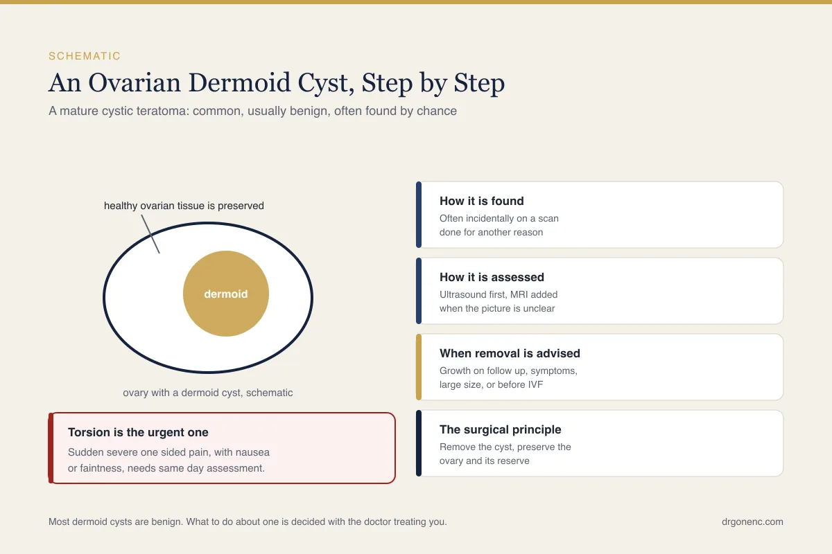
Ovarian torsion is the complication that most often makes a dermoid cyst an emergency, and it is the one most people have not heard of. A dermoid cyst can also burst, which releases its greasy contents into the abdomen and causes sudden severe pain and marked tenderness, and it can bleed into itself. All three present as sudden, severe pelvic pain and all three need assessing the same day, so the list below applies whichever of them is happening. The weight and bulk of the cyst let the ovary pivot on the ligaments that hold it, twisting its blood supply. The ovary then swells and, if the twist is not relieved, its tissue can be lost. This can happen with a cyst of any size, including a small one, and it can happen in someone who had no symptoms at all the day before. It is more likely during pregnancy and after ovarian stimulation, when the ovary is already enlarged and heavier than usual.
- Sudden, severe pain on one side of the lower abdomen or pelvis, often starting abruptly
- Pain that comes in waves, or pain severe enough to wake you or stop you standing straight
- Pain with nausea or vomiting, which is a common and easily dismissed combination
- Pain that begins after exercise, sudden twisting movement or intercourse
- Feeling faint, dizzy, grey, sweaty or breathless, a racing pulse, or collapsing alongside the pain: do not wait and do not travel alone, call the emergency number for your country (112 in Turkey, 999 in the UK) or get straight to an emergency department
- Fever, or feeling generally unwell and getting worse, alongside the pain
- Any sudden one-sided pelvic pain during pregnancy or during or after an IVF cycle
- Pain that is getting steadily worse over hours rather than easing
Apart from faintness, breathlessness or collapse, which need emergency services immediately, any of these needs assessment the same day, through an emergency department or an out of hours gynaecology service, not a routine appointment in a few days. Do not wait to see whether it settles overnight, and do not wait for a scheduled scan. Torsion is diagnosed by examination, ultrasound with Doppler and, when suspicion remains high, by looking directly with a laparoscope, because imaging can be reassuring in a twisted ovary. If torsion is confirmed the usual operation is to untwist the ovary and, where it is practical at the same sitting, remove the cyst. The ovary is normally kept and left to recover even if it looks dark or bruised when the surgeon first sees it, because an ovary that looks badly affected frequently regains normal function; removal is reserved for an ovary that is truly beyond saving. That is a reason to be seen quickly rather than a reason to assume the ovary is already lost.
Why most dermoid cysts are benign, and the uncommon exceptions
A mature cystic teratoma is, by definition, a benign tumour. The tissue inside it is adult tissue that has finished developing, which is what mature means in the pathological description, and it does not invade or spread. This is why these cysts can often be watched rather than removed, and why a woman told she has a dermoid cyst is not being told she has a cancer. Most will never cause her any problem beyond the scan report itself.
There are genuine exceptions, worth stating plainly rather than leaving as an unspoken worry. An immature teratoma is a separate and uncommon malignant germ cell tumour that mainly affects children and young women; it looks different on imaging, often grows faster, often but not always raises tumour markers such as alpha fetoprotein and is treated quite differently. Separately, a long-standing mature dermoid cyst can very occasionally undergo malignant change, most often to a squamous cell carcinoma arising in its skin lining. This is rare, and the risk is concentrated in older and postmenopausal women and in larger cysts, which is why a new or growing cyst of this kind after the menopause is looked at more carefully.
A few rare associations also exist. Some dermoid cysts contain functioning thyroid tissue, called struma ovarii, which can occasionally produce enough thyroid hormone to cause symptoms. A small number contain nervous tissue that triggers an immune reaction against the brain, a condition known as anti NMDA receptor encephalitis, which presents with psychiatric and neurological symptoms rather than pelvic ones. Both are uncommon enough that they are not a reason to remove a quiet cyst, but they are reasons why unexplained symptoms in someone with a known dermoid cyst should be mentioned to her doctor.
When removal is advised and when watchful waiting is reasonable
There is no single size at which every dermoid cyst must come out. The decision weighs several things at once: how large the cyst is and whether it is growing, whether it is causing symptoms, how confident the imaging is that it is benign, the woman's age and menopausal status, whether she is pregnant or planning pregnancy, how much normal ovarian tissue remains around the cyst, and whether there has already been an episode suggestive of torsion. Two women with cysts of identical size can reasonably be advised differently.
- Persistent pain, pressure or bloating that the cyst plausibly explains
- A cyst that is clearly enlarging on repeat scans over months
- Larger cysts. There is no agreed threshold for a dermoid cyst specifically, and the published size figures you may come across, such as the RCOG's 5 cm and 7 cm, apply to simple fluid cysts rather than to this type. In practice many surgeons begin discussing removal somewhere around 5 cm and argue for it more strongly as a cyst approaches 8 to 10 cm, but this is surgical custom rather than a rule, and size is only one input
- Imaging features that are not typical of a benign dermoid cyst, or a mass whose origin is unclear
- A previous episode of torsion, or symptoms strongly suggesting one
- A new or growing cyst of this kind after the menopause
- A cyst discovered before planned ovarian stimulation, where torsion risk and access to follicles both need thinking about
Where none of those applies, observation is a legitimate choice, and published guidance genuinely differs here rather than pointing to one correct interval. Some bodies frame surveillance as a repeat scan after a few months, then less often if the cyst is stable, while others emphasise individual discussion and set no fixed schedule at all; NHS, RCOG and ACOG patient material all describe watchful waiting for benign ovarian cysts without agreeing on the detail. What matters is that someone has taken ownership of the follow-up and that you know when the next scan is due. Ask your own gynaecologist to write the plan down.
The operation, and the principle that protects ovarian tissue
When a dermoid cyst is removed in a woman who may want to conceive, the aim is almost always an ovarian cystectomy performed laparoscopically: the cyst is separated from the ovary and taken out, and the healthy ovarian tissue around it is left behind to carry on working. The ovary is opened along a planned line, the plane between the cyst wall and the ovarian cortex is found, and the cyst is peeled away as intact as possible. Removing the whole lining matters, because tissue left behind is tissue that can regrow.
| Point of comparison | Ovarian cystectomy | Removal of the ovary (oophorectomy) |
|---|---|---|
| What is taken out | The cyst and its lining only | The whole ovary, with the cyst inside it |
| Effect on that ovary | Ovary is kept and continues to ovulate and produce hormones | That ovary no longer contributes eggs or hormones |
| Effect on ovarian reserve | Some loss of surrounding cortex is unavoidable, and careful technique limits it | Reserve now depends on the remaining ovary alone |
| Usually considered when | Fertility matters, the cyst is benign-appearing, and normal ovarian tissue can be identified | The ovary is unsalvageable, almost no normal tissue remains, after the menopause, or when malignancy is suspected |
| Can the cyst return in that ovary | Yes, a new cyst can form in the preserved tissue | No, although the other ovary remains capable of forming one |
| Usual approach | Laparoscopy in most cases | Laparoscopy or open surgery depending on size and the clinical picture |
Two technical points have a direct bearing on fertility and are reasonable to ask about beforehand. The first is haemostasis: heat used freely on the raw ovarian surface damages the follicles sitting just beneath it, so surgeons tend to use fine sutures or sparing, targeted coagulation instead. The second is spillage. Sebaceous material released into the abdomen can irritate the peritoneum, so the cyst is handled gently, often placed in a retrieval bag, and the pelvis washed out thoroughly. If your ovarian reserve is already a concern, for example with a low AMH result, say so before surgery is planned, because it changes the conversation.
Recurrence, and what happens to the other ovary
A dermoid cyst can come back in the same ovary after a cystectomy. That is not usually a sign that the operation was done badly; it reflects the fact that the cyst grows from a germ cell, and the preserved ovary still contains germ cells. Recurrence is uncommon rather than expected, tends to appear years rather than months later, and is more often seen after very large cysts, after cysts that ruptured during removal, where more than one cyst or both ovaries were involved the first time, and in younger women with many reproductive years ahead.
Both ovaries are affected in a meaningful minority of women, and the second cyst is sometimes small enough to be missed on the first scan. For that reason the surgeon will normally inspect the other ovary during the operation and the radiologist will comment on it specifically. A small dermoid cyst found on the opposite side is not automatically removed at the same time, because every operation on an ovary takes healthy tissue with it, and leaving a quiet cyst alone is often the better balance.
After surgery, the practical advice is simple. Attend the follow-up scan you are given, keep the histology report with your own records, and report new persistent one-sided pain rather than assuming it must be scar tissue. Nothing about having had a dermoid cyst removed means you need lifelong scanning, but it does mean a new ovarian finding deserves to be interpreted alongside what happened before.
Dermoid cysts found before or during IVF
A dermoid cyst is quite often picked up at the baseline scan of a fertility assessment, when the ovaries are being looked at closely for the first time. The first question is almost always whether it has to come out before treatment starts, and the honest answer is that it depends on the cyst rather than on a rule. A small, typical, symptomless dermoid cyst sitting at the edge of an ovary with plenty of visible follicles is frequently left where it is, and stimulation proceeds with it in place.
The argument for removing it first rests on two things: a large cyst can make the follicles difficult to see and difficult to reach safely at collection, and an ovary that is already carrying a heavy cyst is more prone to twisting once stimulation has enlarged it further. The argument against is just as real. Cystectomy always removes some ovarian cortex, and in a woman whose reserve is borderline that loss may matter more than the cyst ever would. This is a genuine trade, and it should be discussed with the clinician planning your cycle rather than settled from an article.
If stimulation goes ahead with the cyst in place, the practical concern at egg collection is the needle path. Puncturing a dermoid cyst spills sebaceous material, which can contaminate the follicular fluid being collected and can irritate the peritoneum, so the aspirating needle is deliberately routed around it and some follicles may be left if they cannot be reached cleanly. Your team should know the cyst is there and where it sits before the procedure, and the operator will usually say in advance if access on that side is likely to be limited.
In pregnancy the usual approach is conservative. Most dermoid cysts found on an early scan are watched, because surgery carries its own risks and many cysts cause no trouble at all over nine months. Removal during pregnancy is generally reserved for torsion, for a cyst that is causing significant symptoms, or where the imaging is worrying, and when an operation is needed the second trimester is the period usually preferred. Any sudden one-sided pain in pregnancy, with a known cyst or without one, is a same day problem.
What to ask the doctor looking after you
A dermoid cyst is one of those diagnoses where the explanation given at the time determines how worried someone stays for the next year. It is reasonable to ask for the reasoning behind the plan, and to ask for it in writing. The questions below are the ones that usually change how a patient understands her own situation, and they are worth taking to the appointment on paper so that none of them is forgotten in the room.
- How confident is the scan that this is a benign dermoid cyst, and what features support that
- What is the exact measurement, and when will it next be measured
- Is the other ovary normal on this scan
- Given my age and my plans for pregnancy, what argues for removing it now and what argues for waiting
- If I have surgery, is the plan to remove only the cyst and keep the ovary
- What is the plan if the cyst turns out to be stuck to surrounding tissue or bursts during removal
- What symptoms should bring me back the same day rather than at the next appointment
Nothing in this article replaces that conversation. It is written to make the terminology on your report readable and to make clear which symptoms are urgent, so that the discussion with your own gynaecologist starts from a more useful place. The assessment of your cyst, and any decision about treating it, rests with the doctor who has examined you and reviewed your images.
Related Reading
- Ovarian pain: what causes it, and when it needs urgent assessment
- Low AMH & Diminished Ovarian Reserve
- Endometriosis and Fertility: Symptoms, Diagnosis and Getting Pregnant
- Egg Retrieval: What to Expect Before, During and After the Procedure
- Endometriosis diagnosis: how the condition is tested and confirmed
Sources and references
This article is supported by the following independent, authoritative sources: