Endometriosis diagnosis: how the condition is tested and confirmed
Endometriosis is a condition in which tissue resembling the lining of the uterus grows outside the uterine cavity, most often on the peritoneum lining the pelvis, on the ovaries, and on the ligaments behind the uterus. It is common: the World Health Organization estimates that endometriosis affects around 190 million women of reproductive age worldwide. It is also one of the conditions that women wait longest to have named. Audits and patient surveys across the United Kingdom and Europe describe an interval of several years between the first appointment about period pain and a diagnosis written in the notes, and the World Health Organization puts the average time to diagnosis at between 4 and 12 years.
This article sets out how endometriosis diagnosis actually works in practice: the pattern of symptoms a clinician listens for, what a pelvic examination can and cannot establish, what a transvaginal ultrasound scan shows and what it misses, when magnetic resonance imaging is added, why no blood test rules the condition in or out, and what a laparoscopy still settles. It also explains where NICE, ACOG and ESHRE frame the pathway differently, and how a suspected diagnosis changes fertility planning. Which of these steps applies to you is a judgement for the doctor assessing you.
Why a diagnosis of endometriosis so often arrives late
Several things conspire to delay recognition. Severe period pain is still widely treated as normal, by patients and sometimes by clinicians. The symptoms overlap heavily with irritable bowel syndrome and with bladder conditions, so the first referral is often to the wrong clinic. Hormonal contraception started for heavy bleeding can suppress symptoms enough to postpone the question for years. And there is no simple test: a woman can have extensive disease with entirely normal examination findings and a normal scan, which is often misread as reassurance.
The delay is not only a matter of comfort. Years of poorly controlled pelvic pain affect sleep, education, work and mood, and persistent pain becomes harder to settle the longer it runs. Deep disease involving the bowel, bladder or ureter can progress quietly. In fertility terms the arithmetic is unforgiving, because ovarian reserve falls with age whatever the diagnosis: a woman who finally has her endometriosis named at 38 has a narrower set of choices than the same woman at 30, through no fault of her own.
Naming the condition matters even where the immediate treatment would look similar. A recorded diagnosis changes how future pain is interpreted, opens access to specialist services, prompts earlier fertility assessment, and allows a woman to stop being told that nothing is wrong. It also sets a baseline, so that a change in symptoms years later is read against something rather than against nothing.
Pelvic pain that needs assessment the same day
Almost everything in this article is about the unhurried assessment of pain that has been present for months or years. A small number of presentations are different, because they are time critical rather than merely uncomfortable, and they belong near the beginning rather than at the end. None of them is a reason to assume endometriosis, and none of them should wait for a scan appointment.
- Sudden severe pain on one side that arrived over minutes rather than days, especially with vomiting, which can mean an ovarian cyst has twisted or bled
- Any pelvic or lower abdominal pain when a pregnancy is possible, including before a test has been done, because an ectopic pregnancy is assessed urgently
- Feeling faint, light headed or clammy, or collapsing, with or without any visible bleeding
- Fever with pain, or pain together with an unusual vaginal discharge, which can indicate pelvic infection
- New shoulder tip pain that does not follow your usual cycle and does not follow recent surgery, which can mean blood inside the abdomen irritating the diaphragm
- Breathlessness, chest pain or coughing up blood, whether or not it seems to follow the cycle
- Bleeding that soaks through protection within an hour, or any bleeding with dizziness
- Being unable to pass urine, or a new inability to open the bowels together with vomiting
Attending an emergency department, or an early pregnancy assessment service where one exists, for any of these is not an overreaction. A pregnancy test comes before almost everything else in anyone of reproductive age with new pelvic pain, whatever they believe about their own cycle. The rest of this article is about the slower question of how endometriosis is recognised and confirmed.
The symptom pattern a clinician listens for
Assessment begins with the history, and the single most useful feature is a cyclical pattern. Pain that reliably worsens in the days before and during bleeding, and settles afterwards, points towards tissue that responds to the hormonal cycle. The history also asks where the pain is felt, whether it has changed over years, what relieves it, which painkillers have been tried and at what dose, and whether bowel or bladder symptoms follow the same rhythm. NICE sets out a list of features that should raise suspicion, including infertility alongside any of them.
- Period pain that begins a day or two before bleeding starts and interferes with school, work or sleep
- Pelvic pain that continues beyond the period, or is present most days
- Deep pain during or after intercourse, felt inside rather than at the entrance
- Pain on opening the bowels, or cyclical bowel symptoms that have been labelled as irritable bowel syndrome
- Cyclical bladder symptoms, including pain passing urine or blood in the urine around a period
- Shoulder tip or chest pain that recurs with each period, which can reflect disease near the diaphragm, although new shoulder tip pain outside that pattern, or any breathlessness, chest pain or coughing up blood, needs assessment the same day rather than a routine appointment
- Marked fatigue that tracks the cycle
- Difficulty conceiving, particularly when it sits alongside any of the above
One point deserves emphasis because it is so often misunderstood: the severity of pain does not track the amount of disease. Widespread superficial deposits can be intensely painful, while a sizeable ovarian endometrioma may cause almost nothing and be found incidentally on a scan requested for another reason. Symptoms therefore guide whether to investigate, but they do not predict what will be found, and a stage recorded at surgery does not tell your doctor how much pain you are in.
What a pelvic examination can and cannot establish
NICE advises offering an abdominal and pelvic examination where endometriosis is suspected. The abdomen is palpated for masses and for areas of tenderness. A speculum examination looks at the upper vagina behind the cervix, where deposits are occasionally visible as bluish or reddish spots. A bimanual examination then assesses the size and position of the uterus, whether it moves freely, whether there is tender nodularity in the posterior vaginal fornix, and whether either ovary feels enlarged, tender or fixed in place.
A fixed, retroverted uterus with reduced mobility and nodular thickening behind it is a meaningful finding that makes deep disease considerably more likely. The reverse does not hold. Superficial peritoneal endometriosis cannot be felt at all, so a completely normal examination excludes nothing, and NICE states this explicitly. Findings also depend on the examiner, on how tense the pelvic floor is, on body habitus, and on whether the examination can be completed at all when it is painful.
A pelvic examination should be consented, explained and chaperoned, and it can be declined or deferred. Where a vaginal examination is not appropriate, for example in a young person or someone who has not been sexually active, an abdominal examination is still useful to exclude a palpable mass, and the assessment moves on to imaging by a route that suits the patient. Nothing in the pathway requires an examination that a woman does not want.
What transvaginal ultrasound shows, and what it misses
A transvaginal ultrasound scan is usually the first imaging test. The probe sits close to the pelvic organs, which gives good resolution over a short distance. In experienced hands it usually identifies an ovarian endometrioma, typically as a rounded cyst with uniform low level, ground glass internal echoes. It can show deep nodules in the uterosacral ligaments, the rectovaginal septum, the bowel wall and the bladder, measured in mm. It also picks up fibroids, hydrosalpinx and the features of adenomyosis, which frequently coexists.
A good scan is more than a picture of the ovaries. An experienced operator looks for so called soft markers: ovaries that are tethered and will not slide, ovaries drawn together behind the uterus, site specific tenderness as the probe passes over a particular point, and a negative sliding sign where the front of the rectum no longer glides against the back of the uterus, which suggests the pouch of Douglas is obliterated by adhesions. Structured protocols and operator experience change what a scan detects more than the machine does.
What ultrasound cannot do is show superficial peritoneal disease or fine adhesions, and these account for a great deal of symptomatic endometriosis. A normal transvaginal scan therefore reduces the likelihood of endometrioma and deep disease, but it does not exclude endometriosis, and it should not end the conversation. Where a vaginal probe is not suitable, a transabdominal scan with a full bladder, or in some units a transrectal scan, gives a less detailed but still useful view.
When MRI is added to the picture
Pelvic magnetic resonance imaging is not usually the first test, because a transvaginal ultrasound scan is offered first and can be organised in primary care. Its particular strength is detecting and then mapping deep disease. Where ultrasound or examination has already suggested deep disease, MRI defines how far a nodule extends into the bowel wall, how close it lies to the ureter, whether the bladder is involved, and how the anatomy is distorted by adhesions. That matters because it determines who needs to be in the operating theatre, which is often a colorectal or urological surgeon alongside the gynaecologist, and what a woman needs to be consented for.
MRI is also used when an adnexal mass is not clearly an endometrioma, and to assess the uterine muscle wall when adenomyosis is suspected. Some units ask for a light bowel preparation or give a medication to quieten bowel movement so the images are sharper. There is no ionising radiation, the scan takes longer than an ultrasound, and the report depends heavily on the radiologist being experienced in this field.
In its 2024 update NICE no longer treats MRI as a test to be held back: recommendation 1.5.9 puts a specialist transvaginal ultrasound scan and a pelvic MRI scan on the same footing for diagnosing deep endometriosis and assessing its extent, and recommendation 1.5.10 asks that either scan be planned and interpreted by someone with specialist expertise in gynaecological imaging. What NICE is clear about is that an unremarkable scan settles nothing: recommendation 1.5.11 advises considering laparoscopy in suspected endometriosis even when the ultrasound or MRI scan was normal. In other words, both imaging tests can come back unremarkable in a woman who genuinely has the condition. That is a limitation of the tests, not evidence about the patient, and it is the single most important thing to understand about negative imaging.
Why there is no blood test that settles the question
Patients often arrive asking for the endometriosis blood test, and the honest answer is that it does not exist. The marker most often raised is CA125, a protein measured in U/mL. It can be elevated in endometriosis, but it also rises with ovulation, with menstruation, with fibroids, with pelvic infection, in early pregnancy, in liver disease and in ovarian cancer. Just as importantly, plenty of women with extensive endometriosis have a CA125 within the laboratory reference range.
NICE therefore advises against using CA125 to diagnose endometriosis, and adds that where a level has already been measured, a raised result may be consistent with the condition but a normal result does not rule it out. The 2022 ESHRE guideline similarly recommends against blood, urine and endometrial biomarkers for diagnosis outside research. Work on microRNA panels, menstrual blood analysis and saliva based signatures continues and is genuinely interesting, but none of it has yet entered mainstream guidance.
Blood tests still have a place in the wider assessment, just not for making this diagnosis. Anti-Mullerian hormone in ng/mL or pmol/L, with FSH and oestradiol in mIU/mL and pg/mL taken in the early follicular phase, help describe ovarian reserve where fertility is a concern. A full blood count is sensible with heavy bleeding, and thyroid function is often checked because its symptoms overlap. Your own doctor decides which of these is worth doing.
The shift away from laparoscopy as the automatic first step
For decades laparoscopy with histological confirmation was treated as the reference standard, and in many services nothing was written in the notes until a surgeon had looked. The consequence of that approach was hidden in the waiting list. A woman with classical cyclical pain had to wait for a theatre slot and accept a general anaesthetic before she could be given a name for her symptoms, and in the meantime she was frequently offered nothing.
Guidance has moved. The 2022 ESHRE endometriosis guideline no longer frames laparoscopy as the diagnostic reference standard, recommending instead that clinical assessment and imaging come first, with laparoscopy reserved for women whose imaging is unremarkable but whose symptoms persist, or for whom empirical medical treatment is unsuitable or has not helped. NICE, in its updated endometriosis guideline, emphasises recognising the pattern early, not excluding the condition on normal imaging, and starting treatment without waiting for surgical confirmation where that is appropriate.
The practical consequence for a patient is twofold. First, you may be given a working diagnosis and offered treatment on the strength of your history and a scan, which is a legitimate pathway rather than a shortcut. Second, if the scan is normal and you are told you do not have endometriosis, that conclusion does not follow from the test, and it is reasonable to ask what the plan is should symptoms continue.
What laparoscopy still settles, and what happens during it
Laparoscopy has lost its automatic place at the front of the pathway, but it still answers questions nothing else can. It is the only way to see superficial peritoneal disease directly, to take a biopsy for histology, to record how extensive the disease is using a staging system such as the revised ASRM score or the #Enzian classification, to assess whether the tubes are open, and to treat what is found in the same anaesthetic rather than at a second operation.
- It is usually a day case under general anaesthesia, with fasting and an escort arranged beforehand
- A small incision at or near the umbilicus admits a telescope, and carbon dioxide gas lifts the abdominal wall away from the organs
- Two or three further incisions of a few mm take the working instruments
- The surgeon inspects the peritoneum, both ovaries, the tubes, the pouch of Douglas, the uterosacral ligaments, the bladder, the surface of the bowel and, where indicated, the diaphragm
- Visible disease can be excised or ablated, adhesions divided, and the wall of an endometrioma removed, in the same sitting if this was consented
- Where fertility is the question, dye may be passed through the cervix to show whether the tubes fill and spill
- Afterwards, shoulder tip discomfort from residual gas is common for a day or two, and most people are back to usual activity within about a week after a diagnostic procedure; pain that instead worsens after the second day, or that comes with fever or vomiting, is not expected and should prompt same day contact with the surgical team
It is not a trivial test. Subtle lesions can be missed even by experienced surgeons, and a biopsy that comes back without endometriosis in it does not undo what was seen, which is why the operation note and the histology report should be read together. The recognised risks include injury to bowel, bladder, ureter or blood vessels, infection, port site hernia and the risks of anaesthesia. The decision rests on whether the result will change management, and that is a conversation with the surgeon who would be operating.
Which investigation answers which question
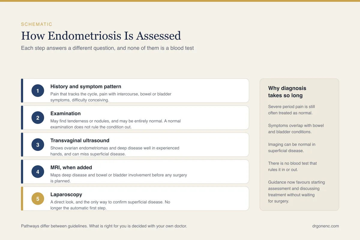
It helps to see the assessment as a sequence of tests with different jobs, rather than as a ladder with one correct answer at the top. Each step narrows the question, and each has a blind spot. The table below sets out what each one can reasonably establish and where it falls silent.
| Step | What it can establish | What it cannot establish |
|---|---|---|
| Clinical history | Whether the pattern is cyclical, how pain affects daily life, which treatments have failed, whether fertility is affected | Where the disease is, how extensive it is, whether another condition explains the symptoms |
| Abdominal and pelvic examination | Palpable masses, tender nodularity behind the uterus, a fixed or immobile uterus, visible deposits in the upper vagina | Superficial peritoneal disease, anything that cannot be reached by a hand, the extent of deep disease |
| Transvaginal ultrasound | Ovarian endometrioma, deep nodules in the ligaments, bowel wall or bladder, obliteration of the pouch of Douglas, adenomyosis, hydrosalpinx, fibroids | Superficial peritoneal deposits, fine adhesions; it cannot exclude endometriosis when normal |
| Pelvic MRI | How far deep disease extends, involvement of bowel, bladder or ureter, distorted anatomy for surgical planning, indeterminate adnexal masses | Superficial disease; it is not usually the first test requested, and a normal scan cannot exclude the condition |
| Blood tests, including CA125 | Ovarian reserve markers, anaemia from heavy bleeding, thyroid function, other causes to consider | The presence or absence of endometriosis; CA125 neither confirms nor excludes it |
| Laparoscopy with biopsy | Direct view of superficial and deep disease, histology, staging, adhesions, tubal patency, treatment in the same procedure | How much pain a woman will have, future progression; very subtle lesions may still be overlooked |
Read the table in both directions. A test that comes back normal has only excluded what that test is capable of seeing, and the column on the right is the part most often left out of a results conversation. If you are told an investigation was normal, it is fair to ask which of these two columns the result belongs in.
Where NICE, ACOG and ESHRE frame the pathway differently
The three bodies agree on more than they differ. All three accept that suspicion should begin with symptoms, that imaging belongs early in the pathway, that CA125 is not a diagnostic test, that normal imaging does not exclude the condition, and that medical treatment for pain can reasonably be started before any surgical confirmation. Where they part company is on what counts as a diagnosis and on how much weight surgery still carries.
NICE, in NG73, organises the pathway around recognition in primary care, abdominal and pelvic examination, transvaginal ultrasound, and referral, while retaining laparoscopy as something to consider when imaging is unremarkable and symptoms persist. ESHRE goes furthest, stating that laparoscopy is no longer the diagnostic reference standard and that a diagnosis made on clinical assessment with imaging is acceptable. ACOG supports starting medical treatment on clinical grounds, and its patient material says you can begin treatment before any imaging, but it still states that the only way to confirm the diagnosis is a diagnostic laparoscopy, with a biopsy taken only in some cases, while being equally clear that no one is required to have that operation.
This is not an academic distinction for anyone moving between health systems. In one country a clinician may record suspected endometriosis and treat it; in another the same woman may be told she cannot be said to have it until a surgeon has looked. Both positions are defensible and both are published. The useful question at your own appointment is which framework your clinician is working within, and what, in practical terms, would change if the label were applied.
How a suspected diagnosis changes fertility planning
Once endometriosis is suspected, fertility assessment tends to move earlier rather than later, because delay carries consequences of its own. That assessment usually means ovarian reserve markers, an antral follicle count on ultrasound, a semen analysis for the partner, and a view on tubal status. Where endometriomas are present, or where previous ovarian surgery has been performed, reserve may already be reduced, and that information changes the pace of planning more than the stage of the disease does.
Several decisions then become specific. Whether to operate on an endometrioma before assisted conception is genuinely contested, because excising a cyst wall removes disease but can also remove surrounding ovarian tissue, so some units prefer to proceed to treatment and leave the cyst alone. Where surgery on both ovaries is planned, discussing egg freezing beforehand is reasonable. Adhesions or a damaged tube may redirect the plan altogether, and this is discussed further in the article on endometriosis and fertility.
The diagnosis also matters for anyone already carrying the label of unexplained infertility, since that label is only as good as the tests behind it, and superficial endometriosis is invisible to all of them. Where the uterine cavity itself is in question, for example after repeated implantation failure, a hysteroscopy answers a different question and is not a test for endometriosis. Which of these steps is worth taking, and in what order, belongs to the doctor reviewing your results.
Who decides what, and when to go back
There is no single correct order for these investigations. The sequence that suits one woman depends on her symptoms, her age, whether she is trying to conceive, whether deep disease is suspected, and how much experience in endometriosis imaging is available locally. Evaluation is individual, and the decision about which tests to request, when to treat on clinical grounds and when to look surgically belongs to the physician who has examined you and read your scans.
Where you are referred matters as much as when. NICE directs anyone with a suspected or confirmed endometrioma, with deep endometriosis involving the bowel, bladder or ureter, or with endometriosis outside the pelvic cavity, to a specialist endometriosis service, meaning an endometriosis centre with a multidisciplinary team, rather than to general gynaecology, and it directs young women and young people aged 17 and under to a paediatric and adolescent gynaecology service or to such a centre. It also asks that specialist transvaginal ultrasound scans and pelvic MRI scans be planned and interpreted by a healthcare professional with specialist expertise in gynaecological imaging. It is entirely reasonable to ask which kind of service you have been referred to, and who will be reading your scan. Whichever service holds your care, go back to it rather than waiting for a scheduled review if any of the following applies.
- Pain that is not controlled by the current plan, or that is steadily worsening
- Bleeding from the bowel or blood in the urine that follows the cycle
- New, severe pelvic pain, particularly with vomiting, fever or faintness, or any pelvic pain where a pregnancy is possible, all of which need assessment the same day, as set out near the start of this article, rather than at the next appointment
- Pain that has begun to prevent work, study or sleep
- Difficulty conceiving after the interval your own clinician set out
- Any result described as normal where your symptoms have not changed
Keeping a simple record helps more than most patients expect. A few months of notes on where the pain is, how it relates to bleeding, what medication was taken and at what dose, and how the pain affected the day gives a clinician something concrete to work from and makes a cyclical pattern visible that memory alone tends to blur. Take it with you, along with copies of any previous scan reports and operation notes.
Related Reading
- Endometriosis and Fertility: Symptoms, Diagnosis and Getting Pregnant
- Adenomyosis vs Endometriosis: Differences, Symptoms, Diagnosis and Fertility
- Hysteroscopy: what to expect before, during and after the procedure
- Unexplained Infertility: Meaning and Options
- Why Does IVF Fail? Common Reasons and What to Do Next
- Ovarian dermoid cyst explained: what it is, how it is found and when it needs treatment
Sources and references
This article is supported by the following independent, authoritative sources: