Retroverted uterus: what a backward tilt means for fertility and pregnancy

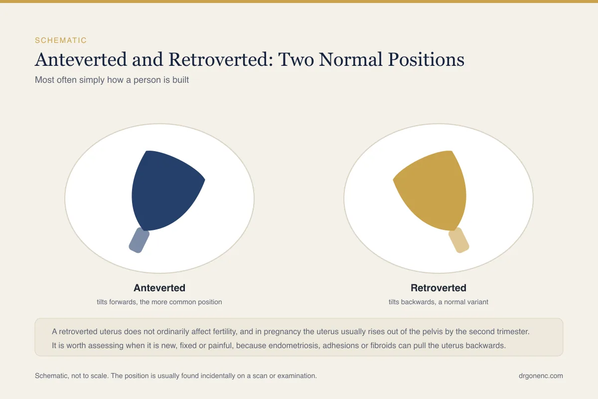
Most people learn that they have a retroverted uterus from a single line on a scan report, or from a sentence said in passing during an examination. The womb, instead of leaning forwards over the bladder, is angled backwards towards the spine and the rectum. The words used vary: retroverted, retroflexed, tilted, tipped, or simply backward. None of them is a diagnosis. All of them describe the direction in which a normal organ happens to sit, and for a sizeable minority of women that direction is backwards from the day their anatomy finished developing.
This article explains what the term means, how it is usually found, and then answers the two questions that bring almost everyone to the subject: whether a backward tilt affects the chance of conceiving, and whether it affects pregnancy. It also covers the situation that matters more clinically, which is when a uterus has been pulled backwards and held there by endometriosis, adhesions or fibroids rather than simply being built that way, and the symptoms that would raise that possibility. Practical sections follow on smear tests, scans and embryo transfer, alongside a clearly marked section on the findings that need assessment the same day.
What a retroverted uterus actually means
The uterus sits in the middle of the pelvis, between the bladder in front and the rectum behind, suspended by ligaments rather than fixed to bone. In the commoner arrangement it leans forwards, so that the upper part rests over the bladder. In a retroverted uterus it leans the other way, with the upper part angled back towards the lower spine. That is the whole of it. The organ itself, its lining, its blood supply and its connection to the fallopian tubes are no different.
Two words tend to appear together on reports and are often used loosely. Version describes the angle between the cervix and the vagina, so a retroverted uterus is one whose whole body is angled backwards at the level of the cervix. Flexion describes the angle between the body of the uterus and the cervix, so a retroflexed uterus is one whose body is bent backwards on its own cervix. Many women are described as retroverted and retroflexed together. For a patient, the distinction almost never changes what happens next.
It also helps to know that uterine position is not fixed the way a bone is fixed. The ligaments that hold the uterus allow it to move, so the angle shifts with a full or empty bladder, with a loaded bowel, with posture, and markedly with pregnancy. A uterus recorded as retroverted on one scan can be recorded differently on another. If you have read about the forward leaning arrangement, the companion article on an anteverted uterus covers the same anatomy from the other direction.
How common it is, and why the uterus tilts the way it does
The patient encyclopaedia of the United States National Library of Medicine records that retroversion of the uterus is common and that in most cases it is a normal finding rather than a disease. Forward tilting is the arrangement found in the majority of women, but backward tilting is common enough that any clinician who examines pelvises sees it regularly and records it without comment. Describing it as abnormal would be inaccurate, and that is why no treatment follows automatically from the finding.
For many women the explanation is simply how they were built. The uterus settled into a backward angle during development and has stayed there since adolescence, with no event to explain it and nothing to find. Natural variation in the length and laxity of the supporting ligaments accounts for a good deal of it. Position can also change later in life: the ligaments stretch during pregnancy and may not return to exactly their previous tension afterwards, so a uterus can be anteverted before a first baby and retroverted after.
It is worth saying clearly what does not cause it, because the internet is unhelpful on this point. A retroverted uterus is not caused by posture, by heavy lifting, by exercise, by using tampons, by a particular kind of sexual activity, or by anything a woman did or failed to do. It is not a consequence of being thin or heavy. If a uterus has been pulled backwards by disease, that is a separate situation covered later in this article, and it comes with symptoms rather than being a silent finding.
How a retroverted uterus is usually found
Nearly always, it is found by accident. A woman has a pelvic ultrasound for something else entirely, a check of the ovaries, an early pregnancy scan, an assessment of heavy periods, and the report includes a descriptive line about uterine position alongside the measurements. Sonographers record it because it tells the next person reading the report how the images were obtained, not because it is a finding that needs acting on.
The other way it comes to light is on examination. During a bimanual examination the examiner places a hand on the abdomen and fingers in the vagina, and feels the body of the uterus. If it cannot be felt in front and is instead felt behind the cervix, the uterus is retroverted. The same examination gives two further pieces of information that matter much more than the tilt itself: whether the uterus moves freely when it is lifted, and whether moving it causes pain.
Because the term is a description rather than a diagnosis, nothing needs to happen when it appears on a report in isolation. There is no procedure to reposition a uterus that is working normally, no exercise programme that changes the angle, and no reason to repeat a scan to check it. The questions that do deserve an answer are whether the uterus is mobile, whether it is tender, and whether the rest of the scan was normal. Those are reasonable things to ask the clinician who ordered the scan.
Does a retroverted uterus affect fertility?
Ordinarily it does not, and that deserves saying plainly rather than hedging. Conception does not depend on the uterus pointing in a particular direction. After intercourse, semen pools in the upper vagina around the cervix, and sperm move into the cervical canal under their own power through cervical mucus, which around ovulation becomes thin and receptive. From there they travel through the uterine cavity and into the fallopian tube, where fertilisation takes place. None of that journey is driven by gravity or determined by the angle of the organ.
This is reflected in how fertility is actually investigated. NICE guidance on fertility problems concentrates on whether ovulation is happening, whether the fallopian tubes are open, what the semen analysis shows, and whether the uterine cavity and endometrial lining are normal. Uterine version does not appear in that assessment as a cause of infertility, and a report that says retroverted, with everything else normal, does not change the plan.
Where a backward tilt becomes relevant to fertility is when it is a clue to something else. Endometriosis, adhesions from previous surgery or infection, and large fibroids can all pull the uterus backwards, and each of those can affect fertility in its own right. So the useful question is not whether the uterus is tilted but why, and whether it still moves. A mobile retroverted uterus in a woman with ordinary periods is anatomy. A fixed, tender one is a finding that needs explaining.
Two related beliefs are worth addressing. Particular positions during intercourse, and lying with the hips raised afterwards, are often recommended on forums for women with a tilted uterus. There is no good evidence that either changes the chance of conceiving, and the reasoning behind them misunderstands how sperm travel. Couples trying to conceive gain far more from understanding the timing of the fertile window than from adjusting for anatomy that was never an obstacle.
Retroverted uterus in pregnancy
In early pregnancy the main practical consequence is on scanning rather than on the pregnancy. A backward-tilted uterus places the gestational sac further from the usual line of sight, so the sonographer may need to angle the probe differently, press a little more, or take longer to obtain clear images and a reliable measurement. This can make an early scan feel slower and less reassuring than expected. It does not mean anything is wrong, and it is not a reason to doubt a dating measurement obtained once the structures are seen clearly.
As the pregnancy grows, the uterus rises out of the pelvis and straightens. By the early second trimester, usually between the twelfth and fourteenth week, the enlarging uterus has lifted into the abdomen and the original tilt has corrected itself. From that point onwards a woman who was retroverted is indistinguishable from a woman who was not, and nothing about antenatal care, delivery or recovery is altered by it. Being retroverted is not an indication for any particular mode of delivery.
Some women are told that a backward tilt explains early pregnancy backache or a feeling of pressure in the rectum. It is plausible, because the growing uterus sits against the sacrum for a few weeks longer, but these symptoms are extremely common in early pregnancy whatever the uterine position, so attributing them is guesswork. Persistent or severe symptoms should be assessed on their own merits rather than explained away by anatomy.
Rarely, a retroverted uterus fails to lift out of the pelvis as the pregnancy grows and becomes trapped below the sacral promontory. This is uncommon, it is described from the second trimester onwards and more often later in pregnancy, and it declares itself with symptoms rather than being a silent finding: increasing pelvic or rectal pressure, difficulty passing urine, and pain. It is the one situation in which a backward tilt in pregnancy genuinely needs urgent attention, and it is set out in the section below.
When a backward tilt is not simply a variant
A uterus can be pulled into a backward position and held there. The commonest reason is endometriosis, where deposits of endometrium-like tissue behind the uterus provoke scarring that tethers the uterus to the rectum and the pelvic side walls. Adhesions following pelvic surgery, a caesarean section, a ruptured appendix or pelvic infection do the same thing mechanically. Fibroids, particularly those growing from the front wall, can tip the uterus backwards by their bulk, and adenomyosis can enlarge and reorient it.
The clinically useful distinction is therefore not retroverted versus anteverted but mobile versus fixed. A uterus that lies backwards but swings freely when the examiner lifts it is anatomy. A uterus that will not move, that hurts when it is moved, or that is accompanied by thickening or nodules felt behind the cervix, is a uterus that something is holding. That is a finding worth investigating, and the investigation is aimed at the underlying condition rather than at the position.
This is also where the distinction between acquired and lifelong position matters. A woman who has always been retroverted, with periods that have not changed, is in a different situation from one whose scan now reports a fixed retroverted uterus after years of worsening period pain. Intrauterine scarring has its own pattern and is covered separately in the article on Asherman syndrome, while the deeper pelvic disease that tethers the uterus is discussed in the article on endometriosis and fertility.
Symptoms that point to a cause rather than a variant
A retroverted uterus that is simply how a woman is built is typically silent. It causes no period pain beyond what she has always had, no change in bleeding, and no difficulty conceiving. When symptoms do cluster around a backward-tilted uterus, they are usually the symptoms of the condition doing the tethering, and they follow recognisable patterns rather than appearing at random. The list below is the one worth raising with a doctor, not because any single item is alarming but because the combination directs the assessment.
- Period pain that has become worse over years, often starting a day or two before bleeding and no longer settling with the pain relief that used to work
- Deep pain during intercourse, felt in the same positions each time and sometimes persisting for hours afterwards
- Pain on opening the bowels or passing urine that is clearly worse during periods
- Pelvic pain present on most days rather than only around the period
- Bleeding that has changed in its own right, becoming heavier, longer or irregular
- Difficulty conceiving after a reasonable period of trying, particularly alongside any of the above
None of these symptoms is specific, and each has explanations that have nothing to do with uterine position. The point of listing them is that they turn a descriptive line on a scan report into a question worth asking. Your own doctor is the person to decide which of them needs examining, which needs imaging, and which needs nothing more than review, because the same symptom carries a different weight at twenty, at thirty-five and after the menopause.
| Feature | Retroversion as a normal variant | Retroversion with an underlying cause |
|---|---|---|
| How it came to light | Noted in passing on a routine scan or examination | Found while investigating pain, abnormal bleeding or difficulty conceiving |
| Movement on examination | Uterus swings freely when the examiner lifts it | Uterus feels fixed and will not move |
| Tenderness | Examination is uncomfortable at most | Pain on moving the uterus or on pressing behind the cervix |
| Period pain | Unchanged from the woman's own usual pattern, or absent | Progressively worse over years, often starting before the bleeding |
| Pain during intercourse | Occasional deep pressure at certain angles | Repeated deep pain, reproducible, sometimes lasting afterwards |
| Rest of the scan | Normal ovaries, normal lining, no fluid | Endometriomas, fibroids, adenomyosis, or an ovary fixed behind the uterus |
| What follows | Nothing; the word is descriptive | Assessment of the underlying condition, planned with your own doctor |
Findings that need assessment the same day
Almost everything on this page is ordinary anatomy that needs no action at all. A small number of situations are different, and they are placed here rather than at the end because the interval between a symptom starting and being assessed is what changes the outcome. With one exception, a uterus that stays trapped in the pelvis as a pregnancy grows, nothing about having a retroverted uterus makes any of these more likely in itself. Women with a backward-tilted uterus sometimes delay seeking help because they have been told their anatomy explains their symptoms, and for acute pain, heavy bleeding, fever or difficulty passing urine it never does.
- Sudden severe pelvic or lower abdominal pain, whether or not there is bleeding: this needs assessing now, not at a routine appointment
- One-sided lower abdominal pain in anyone who could be pregnant, with or without a positive test, because of the possibility of an ectopic pregnancy
- Shoulder tip pain, faintness, dizziness or collapse alongside abdominal pain or bleeding: go to an emergency department or call the local emergency number immediately
- Inability to pass urine, or passing only small amounts with increasing pelvic and rectal pressure, at any stage of pregnancy from the second trimester onwards, which can indicate a uterus trapped in the pelvis and needs urgent assessment the same day
- Heavy vaginal bleeding, for example soaking through a pad within an hour and continuing at that rate, passing large clots, or bleeding with light-headedness, breathlessness or palpitations
- Fever with pelvic pain and vaginal discharge, which can indicate pelvic infection and needs treatment prescribed promptly rather than waiting
- Any bleeding in pregnancy, which should be assessed the same day whatever the uterine position
If any of these apply, seek assessment straight away and do not postpone it in order to read further or to wait for a scheduled appointment. A retroverted uterus is never a reason to wait with acute pain, urinary retention or heavy bleeding, and the one pregnancy situation in which the tilt itself does matter, a uterus trapped in the pelvis, is also one that needs assessing the same day. Treating anatomy as a reassuring explanation is the mistake this section exists to prevent.
Smear tests when the cervix points the other way
This is one of the few places where uterine position has a genuine practical consequence. If the body of the uterus tilts backwards, the cervix tends to point forwards and upwards towards the pubic bone rather than straight down the vagina. The person taking a cervical sample therefore has to angle the speculum differently to bring the cervix into view, and may ask you to shift your hips, place your fists under your buttocks, or change position. It is useful to mention at the start that you have been told your uterus is retroverted.
What changes is the technique, not what the test achieves. A sample taken from a forward-pointing cervix is as good as any other, and the laboratory result means the same thing. If sampling has been difficult or uncomfortable before, saying so allows adjustments, such as a different size of speculum or a slower approach, and there is no reason to accept repeated distress in silence. Persistent difficulty obtaining a sample is itself a reason to be seen by someone more experienced rather than to avoid screening.
It is also worth knowing that cervical screening programmes themselves differ between countries, and this has nothing to do with anatomy. The NHS programme in the United Kingdom invites everyone with a cervix aged 25 to 64 and now screens every five years when a high-risk human papillomavirus test is negative, with earlier recall if it is positive, while American guidance from ACOG begins at 21, with cytology every three years from 21 to 29 and then human papillomavirus testing or co-testing every five years from 30 to 65. Both approaches are considered sound by the bodies that publish them. Follow the programme that applies where you live, and ask your own clinician when your next test is due.
Scans, and what the angle does and does not change
On a transvaginal scan the probe is angled towards the back of the pelvis rather than forwards, which is a routine adjustment for any sonographer. The images obtained are perfectly diagnostic, although a sharply retroflexed uterus can appear foreshortened in some planes, so the operator may take extra views to measure the cavity and the lining in a true long axis. If a measurement of endometrial thickness in millimetres is needed, this matters, because an oblique plane can make the lining look thicker or thinner than it is.
On a transabdominal scan, which is performed through the lower abdomen with a full bladder, a backward-tilted uterus can be harder to see because the upper part of the organ lies behind loops of bowel. For this reason the transvaginal approach is often the more informative one in retroversion, and being offered it is not a sign that anything concerning has been found. A sonographer may also ask you to empty your bladder part way through, because bladder filling changes the angle.
None of this changes what a scan can tell you. The ovaries, the lining, the myometrium, fibroids, polyps, free fluid and early pregnancy are all assessed in exactly the same way. Where a question remains unanswered because of the angle, the next step is a different window or a different modality, decided by the clinician who requested the scan. Structural questions about the shape of the cavity itself, as opposed to the direction it leans, belong to a different subject altogether, that of congenital uterine anomalies, and are assessed with dedicated imaging rather than inferred from uterine position.
Embryo transfer and other procedures through a backward-tilted uterus
Any procedure that passes an instrument through the cervix into the uterine cavity has to follow the curve of the canal, and in a retroverted uterus that curve runs the opposite way to the usual one. This applies to embryo transfer, to fitting an intrauterine device, to endometrial biopsy and to hysteroscopy. Teams anticipate it rather than discover it: uterine position is noted beforehand, often at a preliminary mock or trial transfer, so that the approach is already planned when the day arrives.
The practical adjustments are ordinary ones. Ultrasound guidance shows the catheter tip and the direction of the cavity in real time. A catheter with a different shape, or a malleable one that can be curved the other way, may be chosen. Gentle traction on the cervix can straighten the angle, and bladder filling alters it too, so the clinician may ask for a full bladder in some circumstances and an empty one in others. Which of these is used is a judgement for the person performing the procedure.
The important point is that the angle can make a procedure technically different without changing what it achieves. Uterine version is not among the factors fertility specialists weigh when they discuss the likely course of treatment, and your own clinician is the person to set out what is expected in your case. If a previous transfer or coil fitting was difficult, that is worth mentioning in advance, because it allows the approach to be planned rather than improvised.
What to ask, and what assessment involves
If you have been told you have a retroverted uterus and have no symptoms, the honest answer is that there is nothing to do and nothing to follow up. Three questions are still reasonable to ask the clinician who arranged the scan: does the uterus move freely, was it tender on examination, and was everything else on the scan normal. An answer of yes, no and yes closes the subject, and no further imaging or review is needed for the position alone.
If symptoms are present, assessment starts with the history rather than the scan. A doctor will want to know how period pain has changed over years, whether pain during intercourse is deep and reproducible, whether bowel or bladder symptoms track with the cycle, and how long conception has been attempted. Examination then looks for mobility, tenderness and nodularity. Pelvic ultrasound follows, and depending on what is found, magnetic resonance imaging or laparoscopy may be discussed. Guidance from different bodies varies on how early imaging and surgery should be considered, which is a conversation to have with your own clinician.
The conclusion worth carrying away is a simple one. The phrase on your report describes the direction your uterus happens to lean, in the same way a report might describe the position of your appendix. On its own it predicts nothing about your fertility, nothing about your pregnancy and nothing about your health. What matters is whether it moves, whether it hurts, and what else was seen, and those are questions for the doctor who knows your history rather than for a page on the internet.
Related Reading
- Anteverted Uterus: What It Means, and Whether It Affects Fertility
- Congenital Uterine Anomalies: Bicornuate, Septate, Arcuate, Unicornuate and Didelphys Uterus
- Endometriosis and Fertility: Symptoms, Diagnosis and Getting Pregnant
- Asherman Syndrome (Intrauterine Adhesions) and Fertility
- Endometrial Lining and Implantation: What Matters
- Cramps but no period: what causes it and when it needs assessing
Sources and references
This article is supported by the following independent, authoritative sources: