FSH levels and fertility: what the test shows and what it does not

Most people meet follicle stimulating hormone for the first time on a printed laboratory report, beside a number and a reference interval they have never seen before. FSH is one of the two hormones the pituitary gland uses to speak to the ovaries or the testes, and an early-cycle FSH value forms part of almost every fertility assessment. This article explains what the hormone does, why the timing of the blood sample matters so much, and how a result is read alongside everything else.
Two points are worth making before any figures appear. Reference intervals differ between laboratories, because different analysers and different assay standards return different numbers for the same sample, so the interval printed on your own report is the one that applies to you. And a single FSH value is a measurement rather than a diagnosis. It acquires meaning only next to your age, your cycle history, an ultrasound scan and the other hormones drawn on the same morning.
What follicle stimulating hormone is and what it does
FSH is a glycoprotein hormone released by the anterior pituitary gland at the base of the brain, under instruction from the hypothalamus. In women it acts on the granulosa cells that line the small fluid-filled follicles in the ovary, encouraging them to grow and to convert androgens into oestrogen. In men the same hormone acts on the supporting cells of the testis, sustaining the environment in which sperm are produced. Luteinising hormone is its close partner in both sexes.
The system runs on feedback. As follicles grow they release oestradiol and inhibin B, and both signals travel back to the pituitary and damp the output of FSH. That loop explains the single most useful idea in this whole subject: when fewer small follicles remain, less feedback reaches the pituitary, and the pituitary responds by pushing out more FSH. A raised FSH is therefore an indirect report on the ovary rather than a direct count of anything.
One source of confusion is worth clearing up early. FSH is both something measured and something given. The stimulation injections used in IVF contain FSH, either recombinant or purified from urine, and their purpose is to override the body's own feedback loop so that several follicles grow in one cycle instead of one. That is a different use of the same hormone from the diagnostic blood test this article is about, and the two are not interpreted against each other: a reference interval for a natural day 2 or 3 sample says nothing about the dose given in a treatment cycle. The drugs themselves are described in our guide to IVF injections and medications.
FSH across the menstrual cycle, and why day 2 or 3 matters
FSH is never a fixed number. It rises in the first days of a cycle to recruit a cohort of follicles, then falls back as the growing follicles produce more oestradiol and inhibin B and one follicle becomes dominant. It rises again briefly in the middle of the cycle alongside the luteinising hormone surge, and sits low through the second half while the corpus luteum is active. For the baseline reading used in fertility assessment, a value drawn without knowing the cycle day is hard to interpret. Where periods are absent or very irregular there is no cycle day to record, and the sample is simply taken when it is convenient, a point ESHRE makes explicitly for the investigation of ovarian insufficiency.
Day 2 or day 3 is chosen because it is the most reproducible moment in the cycle: the pituitary is working with very little ovarian feedback, so the figure reflects how hard the gland has to push to get follicles moving. Day one is the first day of proper bleeding, not spotting. Oestradiol is usually drawn from the same sample, and often luteinising hormone as well, because the three readings only make sense together.
If your cycles are irregular, very long, or absent altogether, there is no reliable day 2 or 3 to aim for, and your doctor will decide when to sample and what else to measure at the same time. The same applies if you are taking hormonal medication, which suppresses the pituitary so that the figure no longer reflects your own baseline. In that situation the gonadotrophins are usually read together with oestradiol, prolactin and thyroid function rather than against a cycle-day interval.
Reference ranges and units: what the numbers on your report mean
FSH is usually reported in milli-international units per millilitre, written mIU/mL. You may also see IU/L, and for FSH the two are numerically the same, so a value of 7 mIU/mL is a value of 7 IU/L. The intervals below are indicative figures of the kind laboratories commonly publish, offered so that a result is less bewildering. They are not thresholds for a decision, and they are not interchangeable with the intervals on your own report. One point is worth knowing before you read the table. Sitting inside a laboratory's usual interval is not the same as being of no interest: a value in the upper single figures can still carry weight in a treatment-planning conversation, read beside your age, the oestradiol drawn with it and the follicle count on a scan. NICE's current fertility guideline, NG257, goes further in one direction and advises against using FSH at all to predict how the ovaries will respond to stimulation or the outcome of assisted conception, recommending AMH or an antral follicle count for that question instead.
| Sample and context | Indicative interval (mIU/mL) | What the figure reflects |
|---|---|---|
| Early follicular phase, day 2 or day 3 | roughly 3 to 10 | The baseline used in fertility assessment, taken before ovarian feedback suppresses the pituitary |
| Mid-cycle, around ovulation | roughly 4 to 22 | The short rise that accompanies the luteinising hormone surge as the egg is released |
| Luteal phase, second half of the cycle | roughly 1 to 9 | A quiet phase in which progesterone and oestrogen from the corpus luteum hold the pituitary down |
| After the menopause | roughly 25 to 135 | A sustained high signal because ovarian feedback has largely ceased |
| Adult men, any day | roughly 1 to 12 | The steady drive to the supporting cells of the testis that maintains sperm production |
Several things explain why the same woman can produce different figures. Assay calibration varies between manufacturers. FSH is released in pulses, so the exact minute of the blood draw matters a little. Cycle-to-cycle variation is real and well recognised, particularly from the late thirties onwards. High-dose biotin supplements can interfere with some immunoassays and should be mentioned to whoever takes the sample. For all these reasons an unexpected value is often simply repeated in a later cycle.
A word on home kits, since they are easy to buy. Over-the-counter FSH tests measure the hormone in urine rather than blood and are designed only to flag a level that is higher than usual, which is why they are sold as menopause indicators. They cannot be used to assess fertility: as MedlinePlus puts it, they cannot accurately tell whether the ovaries are making eggs, and a negative strip is not evidence that reserve is normal. They also give no units, no cycle-day context and no oestradiol alongside, which are the three things that make a blood result interpretable. If a home test prompted you to read this page, the next step is a dated blood sample arranged through your doctor rather than a repeat strip.
What a raised FSH reading can suggest
A raised early-cycle FSH suggests the pituitary is working harder than usual to recruit follicles, most often because the pool of small antral follicles, and the inhibin B and oestradiol feedback it generates, has declined. In broad clinical use, day 2 or 3 values from the upper single figures to about 15 mIU/mL are read as borderline and prompt a closer look, while values above roughly 15 to 20 mIU/mL raise a firmer question about diminished ovarian reserve.
Those figures are conventions rather than fixed rules, and they differ between centres and between assays, which is one reason your own laboratory's interval matters more than any number quoted in an article. A raised FSH on its own, in a woman with regular cycles and no difficulty conceiving, may simply be repeated. One high reading in one cycle settles very little.
There is an important trap in the opposite direction. If early-cycle oestradiol is already high, perhaps from a residual cyst or an early-growing follicle, that oestradiol suppresses FSH and can pull a genuinely raised value down into the reassuring range. This is precisely why the two are measured together. The wider picture of reduced reserve is covered in our article on low AMH and diminished ovarian reserve.
Why a single FSH value is not a diagnosis
Before a result is treated as meaningful, a doctor works through the ordinary reasons a figure may be unreliable. Most of them have nothing to do with the ovary itself, and most are easy to establish from a short conversation about timing and medication. The list below is the sort of check that happens routinely in clinic, and it is worth running through your own circumstances against it before you draw conclusions at home.
- The cycle day the sample was taken, and whether bleeding that day was a true period or spotting.
- Combined hormonal contraception, hormone therapy or other gonadotrophin-suppressing medication taken in the preceding weeks.
- An ovarian cyst or an early dominant follicle producing oestradiol that masks the true baseline.
- High-dose biotin or other supplements that can interfere with immunoassay measurement.
- Recent pregnancy, recent pregnancy loss, or breastfeeding, any of which alter the axis for a time.
- A change of laboratory or analyser between one test and the next, so two numbers are not strictly comparable.
When a result does not fit the clinical picture, the usual response is to repeat it in a later cycle at the same laboratory, with oestradiol alongside it and an ultrasound scan in the same window. Interpretation belongs with the doctor who knows your history, examined you and ordered the test, and a value read in isolation on a screen is not a substitute for that.
FSH, AMH and antral follicle count: three windows on one question
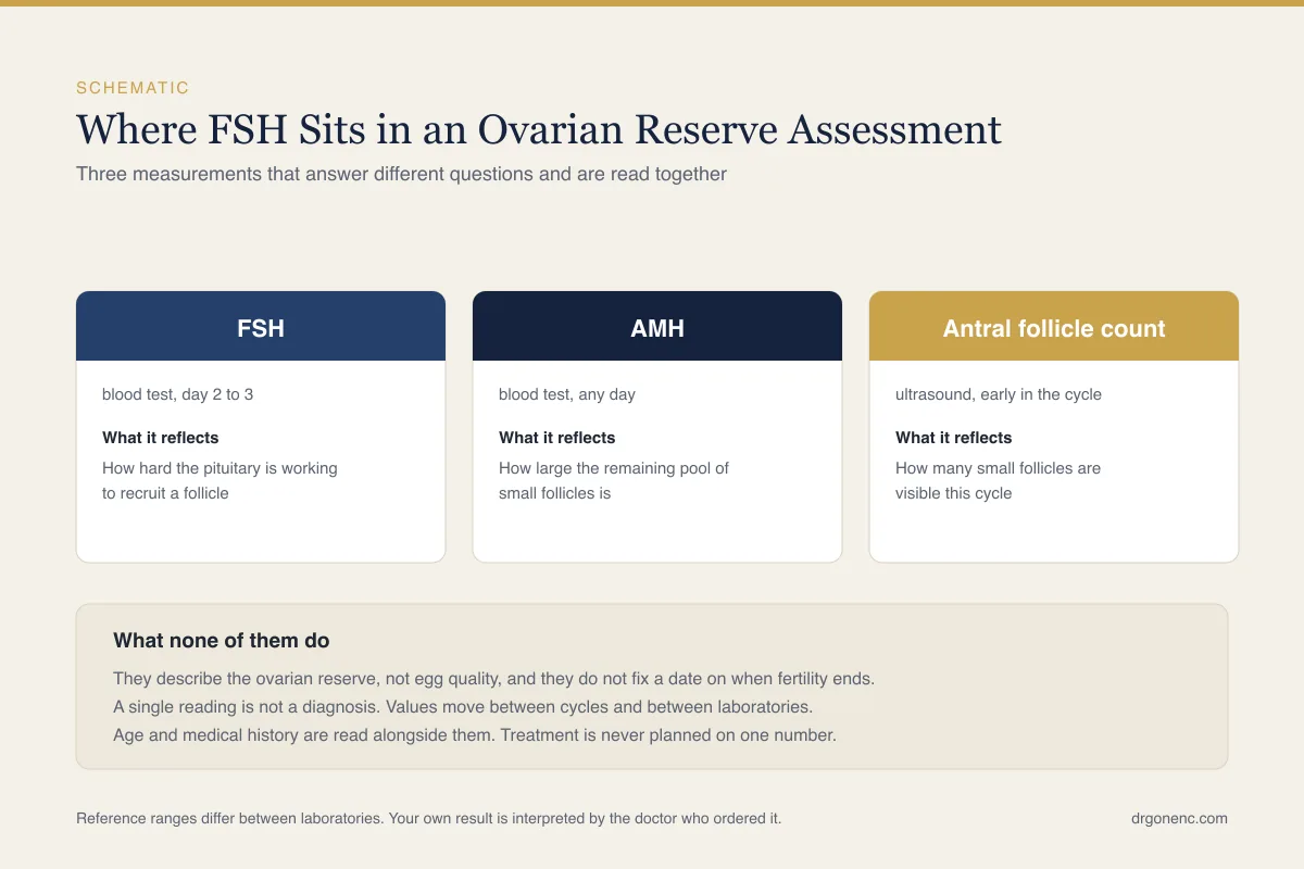
Three tests are commonly used to describe ovarian reserve, and they look at it from different angles rather than competing with one another. Understanding what each one actually measures explains why they sometimes disagree, and why no single one of them is treated as the answer. Guidance differs on how many of them are needed, so your doctor may have one, two or all three in front of them before discussing what reserve means for you.
- AMH is produced by the small growing follicles themselves, so it reports on the size of that pool fairly directly. It can be drawn on any day of the cycle, which makes it practical.
- Antral follicle count is a direct visual count of small follicles on a transvaginal ultrasound scan, usually in the early follicular phase. It depends on image quality and on the experience of the person scanning.
- FSH is a feedback signal from the pituitary, so it tends to shift only once the follicle pool has already fallen appreciably. That makes it a later marker than the other two, and a more variable one from cycle to cycle.
Because of that sequence, AMH and antral follicle count are generally regarded as earlier and more reproducible indicators of reserve, with FSH adding information about how the pituitary is responding. They often point the same way, but disagreement between them is common enough to be an everyday clinical situation rather than a sign that something has gone wrong with the testing. When they do not, the mismatch is a reason to review the timing, repeat a test or scan again, rather than to pick whichever number feels most comforting or most alarming.
How FSH changes with age and towards the menopause
Early-cycle FSH drifts upwards across the reproductive years, slowly through the thirties and more noticeably in the forties, as the follicle pool declines. During perimenopause the behaviour becomes erratic: a woman can record a clearly raised value in one cycle and an ordinary one in the next, because some cycles still recruit follicles effectively. A normal FSH in this phase of life therefore does not rule out the transition, and a single high value does not confirm it.
People often search for an FSH table broken down by age, and it is worth saying plainly that laboratories do not generally publish one. Reference intervals are set by cycle phase and menopausal status, not by year of age, and no professional body has issued age-banded day 2 to 3 cut-offs. What changes with age is the weight a given number carries rather than the number that counts as usual: 9 mIU/mL at 28 and 9 mIU/mL at 42 sit in the same laboratory interval but mean different things for how quickly to investigate, which is why NICE names maternal age itself as the initial predictor to use. The reverse point matters as much: many women with reduced ovarian reserve have an entirely ordinary day 3 FSH, so a normal result does not confirm normal reserve at any age. Any age-banded FSH table you find online is a single clinic's local convention rather than a published standard.
Guidance on when to measure it genuinely differs between bodies. NICE, in its menopause guideline NG23, says perimenopause and menopause can be identified without laboratory tests in otherwise healthy people aged 45 or over who have menopause-associated symptoms, and that serum FSH should be used to confirm menopause only in people aged 40 to 45 with those symptoms including a change in their menstrual cycle, or in people under 40 in whom menopause is suspected. It also advises against an FSH test in anyone using combined oestrogen-and-progestogen contraception or high-dose progestogen. Other organisations and laboratories frame their advice somewhat differently, so your doctor works to local protocol.
Premature ovarian insufficiency, the term ESHRE uses for the condition that is also widely called primary ovarian insufficiency, has thresholds and testing rules of its own, and these vary too. The 2024 ESHRE guideline diagnoses it on disordered cycles, meaning spontaneous absence of periods or irregular periods, for at least four months together with an FSH above 25 IU/L, and it advises repeating the FSH after four to six weeks only where the diagnosis is uncertain, since a second sample can confuse the picture if the first was already clear. NICE asks instead for a raised FSH on two blood samples taken four to six weeks apart and says the diagnosis should not rest on a single blood test. Older sources used a higher cut-off such as 40 IU/L. What all of them agree on is that this is not the same thing as an early menopause and does not mean ovulation has stopped permanently.
Low or unexpected FSH patterns
A low FSH attracts less attention than a high one, which is unfortunate, because in a woman whose periods have stopped it can be just as informative. The pattern matters more than the single value: FSH is read with luteinising hormone, oestradiol, prolactin and thyroid function, and it is the shape of the group that points to where the problem sits in the chain from hypothalamus to pituitary to ovary.
- Low FSH and low luteinising hormone with low oestradiol point towards the hypothalamus or pituitary, for instance in hypothalamic amenorrhoea related to low body weight, heavy training or sustained stress.
- Raised prolactin can suppress the whole axis and produce low gonadotrophins with absent periods, which is why it is usually measured in the same panel.
- Polycystic ovary syndrome more often shows FSH in the normal or low-normal range with luteinising hormone relatively higher, frequently alongside a high AMH and a high follicle count on scan. The raised LH-to-FSH ratio is common rather than universal, and it is not itself a diagnostic criterion, which is why PCOS is not diagnosed from gonadotrophins alone. Some patient-facing sources also list PCOS among the causes of a high FSH, which is a further reason not to read a single gonadotrophin value as a diagnosis.
- Thyroid disorders and recent hormonal treatment can shift gonadotrophin readings without any change in the ovary itself.
The practical conclusion is that absent or very irregular periods deserve investigation whether FSH comes back high, low or unremarkable, because a reassuring number does not explain a missing cycle. A raised prolactin, which is one of the patterns above, is covered separately in our article on high prolactin and fertility.
FSH in men
In men FSH acts on the Sertoli cells of the testis, the supporting cells that nurse developing sperm, so it is a reasonable indicator of how the sperm-producing machinery is being driven. There is no cycle to time around, so the sample may be taken on any day. It is usually requested with luteinising hormone and total testosterone, and the testosterone sample is taken in the morning because that hormone follows a daily rhythm.
Three patterns recur. A raised FSH with a low sperm count suggests the testis itself is not producing sperm effectively, so the pituitary is pushing harder. A normal FSH with no sperm in the ejaculate and normal testicular volume raises the possibility of an obstruction in the ducts rather than a production failure. Low FSH and low luteinising hormone with low testosterone points instead to a hypothalamic or pituitary cause, which is managed quite differently.
FSH does not grade sperm. It describes the signal arriving at the testis, not what the testis delivers, so it never replaces a semen analysis, a physical examination, scrotal ultrasound where indicated, or genetic testing in selected cases. Our guide to semen analysis results explains the parameters a hormone test cannot cover.
What an FSH result does and does not predict
Used properly, FSH helps a clinician with a small set of real questions. It forms part of the overall description of ovarian reserve, although NICE's current fertility guideline advises against using FSH as the predictor of ovarian response and points to AMH or an antral follicle count for that particular question. It flags when evaluation should not be postponed. It helps work out why periods have become irregular or stopped. In men it separates a production problem from a transport problem or a pituitary one.
What it cannot do is tell an individual whether or when she will conceive. It cannot date the menopause. It cannot measure the quality of any particular egg, which tracks far more closely with age than with any hormone assay. Women with clearly raised FSH do conceive, and women with entirely ordinary FSH sometimes do not, which is why outcomes are discussed in words and in context rather than as figures.
If treatment is being considered, the hormone result is one input among several into a plan that also weighs age, cycle history, scan findings, the male assessment and previous attempts. No single figure in that list settles the plan on its own, and which of them carries most weight differs from one person to the next.
Bringing the picture together with your doctor
Assessment of fertility is individual, and the interpretation of any hormone result belongs to the physician who is treating you. If you are bringing an FSH result to a consultation, bring the report itself rather than the number from memory: the units, the laboratory's own reference interval, the cycle day, the oestradiol drawn with it, and a list of any medication or supplements you were taking. Those details are what turn a figure into information.
It is also reasonable to ask what the result changes. A good consultation should be able to say whether the value alters the plan, whether it needs repeating, and what the next test would add. Where guidance differs between professional bodies, as it does over when to measure FSH at all, your doctor will explain which protocol they are following and why it applies to your situation.
Related Reading
- Low AMH & Diminished Ovarian Reserve
- Fertility Testing: How to Know If You Are Fertile (Female and Male)
- Getting Pregnant After 40: Real Chances, Risks and Your Options
- PCOS and Getting Pregnant: Symptoms, Diagnosis and Fertility Options
- How Is IVF Done? A Step-by-Step Guide to the IVF Process
Sources and references
This article is supported by the following independent, authoritative sources:
- MedlinePlus (US National Library of Medicine), Follicle-stimulating hormone (FSH) levels test
- NICE (UK), Fertility problems: assessment and treatment (NG257)
- ESHRE, Guideline on premature ovarian insufficiency (2024)
- NHS (UK), Early menopause
- NICHD (US), How do healthcare providers diagnose POI?
- NICE (UK), Menopause: identification and management (NG23)